Biology Reference
In-Depth Information
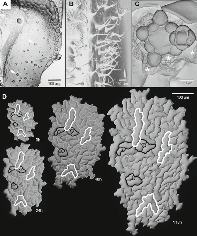
Fig. 1
SEM micrographs showing exemplary casts of various organs of
Anagallis arvensis
(
a
) and
A. thaliana
(
b
-
d
). (
a
) Abaxial surface of a young expanding leaf with grandular trichomes (
arrows
). Note the complex
shape of the leaf in this developmental stage. (
b
) Basal portion of a rosette leaf, whose abaxial surface is
covered by large, branched trichomes. Note how well the complex trichome shapes are reproduced in the cast.
(
c
) Infl orescence shoot apex grown in in vitro culture on agar-solidifi ed medium. The same specimen can be
observed in vivo in CLSM (
see
Chapter
9
). Exemplary places from where older organs (fl ower buds, cauline
leaves) surrounding the apex have been removed are pointed by
asterisks
. (
d
) Sequence of replicas taken from
abaxial leaf surface of
transparent testa glabra1
mutant plant. The time at which replicas were taken is given
in lower right corner of each image. Exemplary cells or cell packets are outlined: in
black
if no cell division took
place or in white if the cells are still dividing. Images shown in (
a
,
d
) were taken with the aid of SEM machine
LEO435VP; (
b
)—Philips XL 30 TMP ESEN; (
c
)—Hitachi S-800
each time, it is convenient to put a small amount of each in
two disposable syringes (10-15 ml volume) and close syringes
by original caps or short injection needles.
Search WWH ::

Custom Search