Biology Reference
In-Depth Information
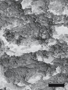
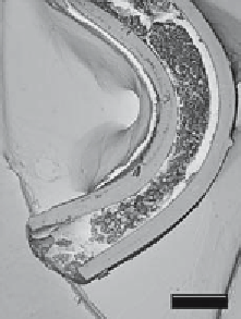
Fig. 3. Scanning electron micrograph of catheter from the bladder of a mouse
infected with
P. mirabilis
Xen 44. Cross-section of catheter at low magnification
(left) shows the lumen of catheter filled with a thick biofilm, and numerous rod-
shaped bacteria embedded within the polymeric substance is clearly seen at higher
magnification (right).
Fig. 4.
In vivo
bioluminescent monitoring of the progression of
P. mirabilis
Xen 44
in the mouse model of catheter-induced urinary track infection (UTI). The ascending
nature of uropathogen from the bladder up the ureters and to the kidney can be
clearly visualized from outside intact live animals using luciferase tag pathogen and
bioluminescence imaging. (
See
Color Plate 8, following p. 46.)


Search WWH ::

Custom Search