Biology Reference
In-Depth Information
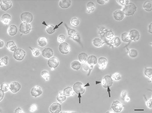
Fig. 2. Human monocyte-derived macrophages (MDMs) infected with the Nine Mile
phase I strain (RSA493) of
Coxiella burnetii
. MDMs in a 24-well plate were infected
for 2 days, then viewed by phase contrast light microscopy using a Nikon TE-2000E
inverted microscope equipped with a CoolSNAP HQ digital camera (Roper Scientific,
Tuscon, AZ). Images were acquired using Metamorph software (Universal Imaging,
Dowingtown, PA) and processed using Adobe Photoshop (Adobe Systems, San Jose,
CA). In macrophages, the replicative vacuole (arrow) enlarges to encompass nearly all
the intracellular space that causes the cells to round up and pushes the phase-bright
nucleus (arrow heads) to the cell periphery. Bar, 25 μm.
and bacterial) DNA is extracted from infected cells in a 24-well plate using an
Ultraclean Microbial DNA Isolation Kit.
2. Remove the culture medium to a 2-mL microfuge tube and centrifuge at 20,000 ×
g
for 15 min to pellet any extracellular bacteria or cells that have detached.
3. To retrieve adherent macrophages, add 300 and 50 μL of microbead and MD1
kit solutions, respectively, directly to well. Pipette up and down to lyse cells and
transfer the lysate to the tube containing the pellet from
step 2
(
see
Note 16
).
4. From this point, follow kit instructions verbatim to obtain purified DNA from
infected cells.
5. Set up reactions to determine the number of
C. burnetii
genome equivalents in
each DNA sample. For each unknown DNA sample, make 80 μL of TaqMan
Universal PCR Master Mix containing 10 μ
M
of dotA-F and dotA-R primers, and
333 n
M
of dotA-probe, as per kit instructions. Mix 10 μL of purified DNA from
step 4
with Master Mix for a final volume of 90 μL. To generate a standard curve
of 10
3
-10
8
dotA copies, add 10 μL of 10-fold dilutions of purified pCR2.1-TOPO
DNA containing the
C. burnetii dotA
gene to 80 μL of Master Mix (
see
Note 17
).
To perform PCR reactions in triplicate, add 3-25 μL aliquots of the 90 μL Master
Search WWH ::

Custom Search