Biology Reference
In-Depth Information
4. Label the samples by immersion into 5 n
M
secondary quantum dot conjugate,
such as goat anti-mouse 655 nm quantum dots, in blocking buffer. Incubate for
30 min at room temperature.
5. Wash the samples at least twice for 15 min in blocking buffer. Wash the samples
two more times in PBS to remove residual protein from the blocking buffer. The
samples are now ready to examine by fluorescence microscopy and to proceed
with processing for TEM or SEM (
see
Subheadings 3.1
and
3.2
).
3.3.3. Imaging by TEM
Quantum dots are readily detectable by TEM in thin section and negatively
stained preparations at voltages commonly used for biological material between
60 and 120 kV. No special requirements are needed for imaging.
3.3.4. Imaging by SEM
Quantum dots are detectable by SEM by topographical contrast with
secondary electron imaging (SEI), compositional contrast with backscattered
electron imaging (BEI), and scanning transmission electron imaging. By SEI,
the quantum dots appear as particles on exposed surfaces that can resemble
normal biological structures. However, the CdSe core and ZnS coating impart
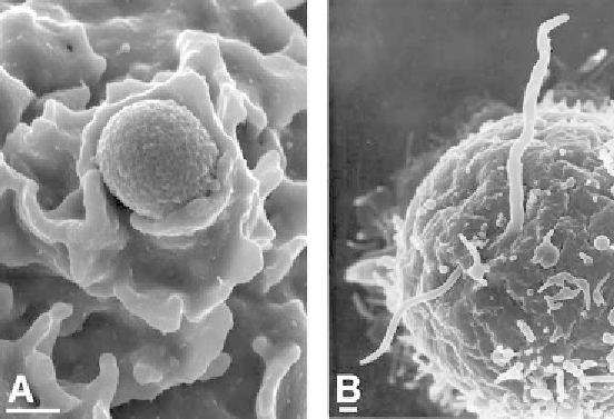
Fig. 2. Scanning electron micrographs showing interactions between bacteria and
host cells.
(A)
Digital image of methicillin-resistant
S. aureus
enwrapped within lamel-
lapodia on the surface of a primary human neutrophil.
(B)
Photographic image of
B. burgdorferi
and a human lymphocyte. The spirochete appears to simultaneously
penetrate and emerge from the B cell, lymphocyte (unpublished data). Bars, 500 nm.
Search WWH ::

Custom Search