Biology Reference
In-Depth Information
Centrosomes as Microtubule-Organizing Centers
The discovery of centrosomes at the end of the nineteenth century is related to the
work of Edouard van Beneden (1846-1910) and Theodor Boveri (1862-1915). In
1883, van Beneden described a new organelle in the middle of the cell, which Boveri
called “centrosome” (1888) and “centriole” (1895). By the end of the nineteenth cen-
tury, they independently developed the hypothesis that centrioles were centers of cell
division (
Heuttner, 1933
).
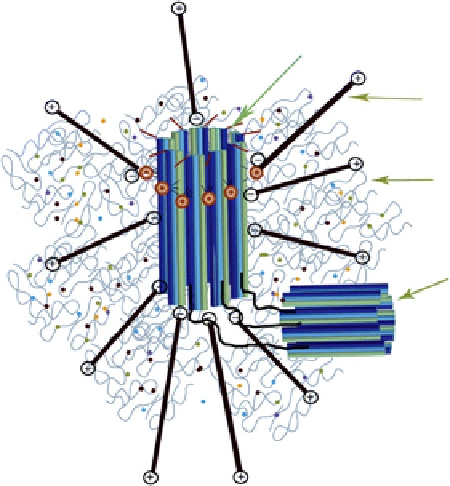
Centrosomes are nonmembranous organelles, which, depending on the position
of the nucleus, are mostly positioned along the middle of the cell, attached to the
nucleus. Centrosomes consist of a spherical body of pericentriolar material (PCM),
proteins embedded on a fibrous lattice. At the middle of the centriole is a pair of cen-
trioles, hollow cylinders measuring about 100-150 nm in diameter and 210-400 nm
in length (
Delattre and Gönczy, 2004
). Centrioles are arranged perpendicularly to
each other and connected through coiled proteins (
Figure 1.21
). In the process of cell
division, centrosomes form the poles of the spindle.
Centrioles are cylindrical structures formed by nine triplet sets of microtubules.
During cell division, the centrosome divides and centrioles migrate to opposite poles,
forming two centrosomes, one for each cell formed. At the start of the transition
from the G1- to the S-phase of the cell cycle, centrioles begin to duplicate; next, each
Mother centriole
Microtubule
PCM
Daughter
centriole
Figure 1.21
A schematic diagram of a typical mammalian centrosome composed of two
centrioles (mother and daughter centriole) surrounded by a meshwork of PCM (
Schatten and
Sun, 2009
).
Search WWH ::

Custom Search