Biology Reference
In-Depth Information
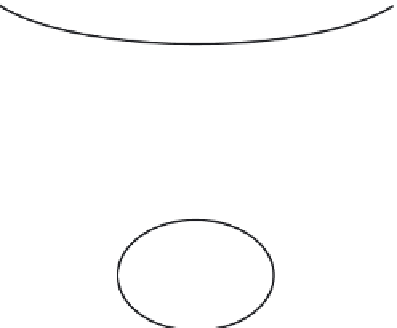
Hypothalamus
Kiss-1 neuron
Kisspeptin
GnRH neuron
Brain
GnRH
Gonadotrope
cell
Acetylation of
histone H3
FSH
Granulosa
cell
Estradiol
Figure 4.5
Neural control of the signal cascade that leads to acetylation of histone H3
and, consequently, induction of FSH-responsive genes and the synthesis of estradiol by
granulosa cells. Note that the epigenetic information necessary for H3 acetylation flows from
hypothalamic neurons, via the FSH-secreting pituitary cells, to granulosa cells of the ovarian
follicle, where it induces FSH-responsive genes and their differentiation and proliferation.
Abbreviations
: FSH, follicle-stimulating hormone; GnRH, gonadotropin-releasing hormone.
form binds to TREs thus preventing the transcription of the TH-regulated genes
(
Moore and Guy, 2005
).
Folliculogenesis is regulated by the pituitary FSH. The hormone binds its spe-
cific receptor in the cell membrane of the granulosa cells, which surround the oocyte.
There it activates the protein kinase A (PKA) transduction pathway that phospho-
rylates and acetylates histone H3. The resulting chromatin remodeling enables tran-
scription of FSH-responsive genes in granulosa cells, inducing their proliferation and
differentiation (
LaVoie, 2005
;
Salvador et al., 2001
). Tracing back the signal cascade
that induces granulosa cell proliferation, differentiation, and estradiol secretion leads
us to a neural source of the information (
Figure 4.5
).
Injection of agonists of receptors for the neurotransmitters dopamine (DA), mus-
carinic acetylcholine (mACh), and glutamate (GLU) caused chromatin remodeling
by inducing a specific modification (phosphorylation) of histone H3 in specific hip-
pocampal neurons. It was concluded that “phosphorylation of histone H3 is coupled
to increased neuronal activity and is directly linked to IEG transcription”. (
Crosio
et al., 2003
).


Search WWH ::

Custom Search