Biology Reference
In-Depth Information
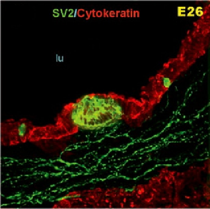
Figure 3.35
Airway epithelium in E26 lung, visualized using antibody against cytokeratin
(red). Corpuscular NEB and two single PNECs (green) localized within epithelial lining.
A rich network of submucosal nerve plexus (green) sends branches toward NEB base. No
apparent neural contacts are seen with single PNEC in this section plane.
Abbreviation
: lu,
airway lumen.
Source
: From
Pan et al. (2004)
.
territory and develops into two endodermal bronchial buds. The Anlagen bifurcate
and evaginate, concomitant with migration of the hindbrain NCCs that colonize the
lung buds, where they differentiate into neurons and glia cells (
Freem et al., 2012
).
On their way to the lung buds, they apparently follow the glial cell line-derived neu-
rotrophic factor (GDNF) as an attractant. The incipient bronchi and epithelial tubules
are encircled by a mesenchyme-derived smooth airway muscle (ASM) and are
closely related to NCCs (
Burns et al., 2008
).
The first lung epithelial cells to differentiate are the pulmonary neuroendocrine cells
(PNECs) at 8 weeks (
Delgadoa et al., 2000; Emanuel et al., 1999
). Shortly thereafter,
clusters of PNECs, known as neuroepithelial bodies (NEBs), appear (
Figure 3.35
).
These neuroendocrine cells are innervated very soon after their differentiation.
They release the neurotransmitter serotonin and bombesin-like peptides (BLPs),
thus inducing the differentiation of various epithelial cell types (
Emanuel et al.,
1999
). Lung vascularization and angiogenesis are also involved in lung development
(
Warburton et al., 2000
), but it is well known that these processes are regulated by
the local innervation that forms from NCCs. The serotonin released by lung neu-
rons is believed to play an important role in lung growth, lung cell proliferation, and
differentiation (
Pan et al., 2006
), and the presence of NEBs (predominantly in the
branching points of bronchi) is believed to reflect its role in lung branching.
Search WWH ::

Custom Search