Environmental Engineering Reference
In-Depth Information
Varnish
White layer
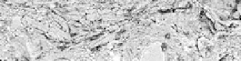
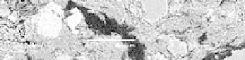
Figure 11.15.
Electron microscopy - backscatter electron pictures showing
the three different layers: preparation layer, white layer and varnish
− The color layer (1-2 mm) had infiltrated the preparation layer and
demonstrated application on the preparation layer when it was still fresh. The white
color came from a talc application, Mg
3
(Si
4
O
10
(OH)
2
); aluminum, silicium and
magnesium were identified by EDX-analysis
(
see Figure 11.21, right-hand side).
The red was composed of hematite and kaolinite, and the black showed poorly
crystallized iron oxide and kaolinite. Organic materials in the color layer were not
analyzed.
− A brown and transparent varnish (15 µm) made of organic material enriched in
saccharides was found. Chromatography analysis was only able to identify a few
components, as the organic materials were old and altered.
11.2.3.2.
Alteration patterns
Next to the south angle of the cathedral, gypsum crystallization (calcium sulfate)
was identified in the adobe wall and under the decorated surfaces (see Figure 11.22,
left). These crystallizations involved a crumbling disintegration under the paint layer
and in some places had completely destroyed the support layer of the decorated
surfaces. Salts were transported by capillary action from the soil to the inside of the
wall and evaporated on the surfaces.
A concrete plate placed against the inside wall might deter the humidity from
evaporating through the ground and push the moisture directly up inside the adobe
wall.