Biomedical Engineering Reference
In-Depth Information
2.3.2 Tertiarystructure
As mentioned previously, a polypeptide's tertiary structure refers to its exact three-dimensional struc-
ture, relating the relative positioning in space of all the polypeptide's constituent atoms to each other.
The tertiary structure of small polypeptides (approximately 200 amino acid residues or less) usu-
ally forms a single discrete structural unit. However, when the three-dimensional structure of many
larger polypeptides is examined, the presence of two or more structural subunits within the polypep-
tide becomes apparent. These are termed domains. Domains, therefore, are (usually) tightly folded
subregions of a single polypeptide, connected to each other by more fl exible or extended regions. As
well as being structurally distinct, domains often serve as independent units of function. Cell surface
receptors, for example, usually contain one or more extracellular domains (some or all of which par-
ticipates in ligand binding), a transmembrane domain (hydrophobic in nature and serving to stabilize
the protein in the membrane) and one or more intracellular domains that play an effector function
(e.g. generation of second messengers). Many therapeutic proteins also display several domains.
Tissue plasminogen activator (tPA), for example (Chapter 12), consists of fi ve such domains.
2.3.3 Higher structure determination
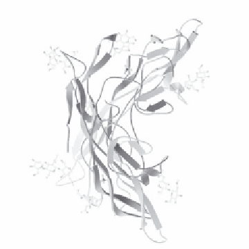
There are three potential methods by which a protein's three-dimensional structure can be visualized:
X-ray diffraction, NMR and electron microscopy. The latter method reveals structural information
at low resolution, giving little or no atomic detail. It is used mainly to obtain the gross three-dimen-
sional shape of very large (multi-polypeptide) proteins, or of protein aggregates such as the outer viral
caspid. X-ray diffraction and NMR are the techniques most widely used to obtain high-resolution
protein structural information, and details of both the principles and practice of these techniques may
be sourced from selected references provided at the end of this chapter. The experimentally deter-
mined three-dimensional structures of some polypeptides are presented in
Figure
2.8.
Figure 2.8
Three-dimensional structure of (a) human interleukin-4, as determined by NMR, and (b) human
follicle-stimulating hormone, as determined by X-ray diffraction. Reproduced from protein data bank (www.
rcsb.org/pdb, molecule ID numbers 1 ITM and 1 FL7 respectively)

Search WWH ::

Custom Search