Biomedical Engineering Reference
In-Depth Information
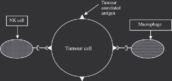
Figure 13.4
Binding of appropriate antibody to tumour-associated antigens marks the tumour cell for de-
struction. This is largely due to the presence of a domain on the antibody F
c
region (see also Box 13.2), which
is recognized and bound by macrophages and NK cells. Therefore, congregation of such cells on the surface of
the tumour is encouraged. This greatly facilitates their cytocidal activity towards the transformed cells
An allied application of radiolabelled anti-tumour monoclonal antibodies is that of diagnostic
imaging (immunoscintigraphy). In this case, the radioisotope employed must be a
-emitter (such
that the radioactivity can penetrate outward through the body for detection purposes). Although
various radioisotopes of iodine have been evaluated, technetium (
99m
Tc) is the one most commonly
employed. It has a γ-ray emission energy that is suffi cient, but a relatively short half life of 6 h (this
minimizes long-term exposure of patient to high-energy
γ
-rays). It can also be generated at nuclear
installations relatively easily and is inexpensive. Equally important, chemical methodologies exist
which facilitate its (stable) coupling to antibody molecules. Direct labelling with
99m
Tc gener a l ly en-
tails initial reduction of antibody disulfi de residues, forming free sulfahydryl (!SH) groups. This
is achieved by incubation with a suitable reducing agent, such as ascorbic acid or sodium dithionite.
A source of
99m
Tc (e.g. Na
99m
TcO
4
) is then reduced separately. Subsequent mixing under nitrogen
gas (to maintain reducing conditions) results in direct linkage of the radioisotope to the antibody.
Upon administration, the anti-tumour
99m
Tc conjugate will congregate at the tumour site. The tumour
can then be visualized using suitable
γ
-ray detection equipment, such as a planar gamma camera.
A number of radiolabelled monoclonal antibodies have been approved as tumour diagnostic
imaging agents (Table 13.2). Carcinoembryonic antigen (CEA)-SCAN, for example, is an antigen-
binding fragment (Fab) of a specifi c murine monoclonal raised against human CEA. As discussed
in detail in Section 13.3.4, CEA is expressed at high levels by some tumours. This is particularly
true of tumours of the gastrointestinal tract, such as carcinomas of the colon or rectum. CEA-
SCAN (non-proprietary name: Arcitumomab) is used to detect these carcinomas. However, CEA
is expressed naturally (all be it at much lower levels) by some non-transformed cells. Therefore,
this antibody fragment is used mostly to image recurrence and/or metastases of histologically
demonstrated carcinoma of the colon or rectum. It is used as an adjunct to standard imaging tech-
niques, such as a computed tomography scan or ultrasonography. Its industrial method of produc-
tion is overviewed in Figure 13.5. The product is administered by i.v. injection, and only relatively
mild side effects are usually noted. These can include nausea, fever, rash and headaches.
Anti-tumour monoclonal antibodies can also be used to deliver toxins to tumour sites. Tox-
ins conjugated to therapeutic antibodies include ricin, pokeweed toxin,
Pseudomonas
toxin and
γ
Search WWH ::

Custom Search