Biomedical Engineering Reference
In-Depth Information
Box 11.3
An overview of the female reproductive cycle
The human female reproductive (ovarian) cycle is initiated and regulated by gonadotrophic
hormones. Day 1 of the cycle is characterized by commencement of menstruation: the dis-
charge of fragments of the endometrium (wall of the womb) from the body, signifying fertiliza-
tion has not occurred in the last cycle. At this stage, plasma levels of FSH and LH are low, but
these begin to increase slowly over the subsequent 10-14 days.
During the fi rst phase of the cycle, a group of follicles (each of which houses an egg) begins
to develop and grow, largely under the infl uence of FSH. Shortly thereafter, a single dominant
follicle normally emerges and the remainder regress. The growing follicle begins to synthesize
oestrogens, which, in turn, trigger a surge in LH secretion at the cycle midpoint (day 14). A
combination of elevated FSH and LH levels (along with additional factors such as prostaglan-
din F
2a
) promotes follicular rupture. This releases the egg cell (ovulation) and converts the
follicle into the progesterone-secreting follicular remnant: the corpus luteum. Release of the
egg marks the end of the fi rst half (follicular phase) of the cycle and the commencement of the
second (luteal) phase.
In the absence of fertilization subsequent to ovulation, the maximum life span of the
corpus luteum is 14 days, during which time it steadily regresses. This, in turn, promotes
slowly decreasing levels of corpus luteum hormones, i.e. oestrogen and progesterone. Pro-
gesterone normally serves to prepare (thicken) the lining of the womb for implantation of
an embryo should fertilization occur. Withdrawal of hormonal support as the corpus luteum
regresses results in the shedding of the endometrial tissue, which marks commencement
of the next cycle. However, if ovulation is followed by fertilization, then the corpus luteum
does not regress but is maintained by hCG, synthesized in the placenta of pregnant females
(Figure 11.B1).
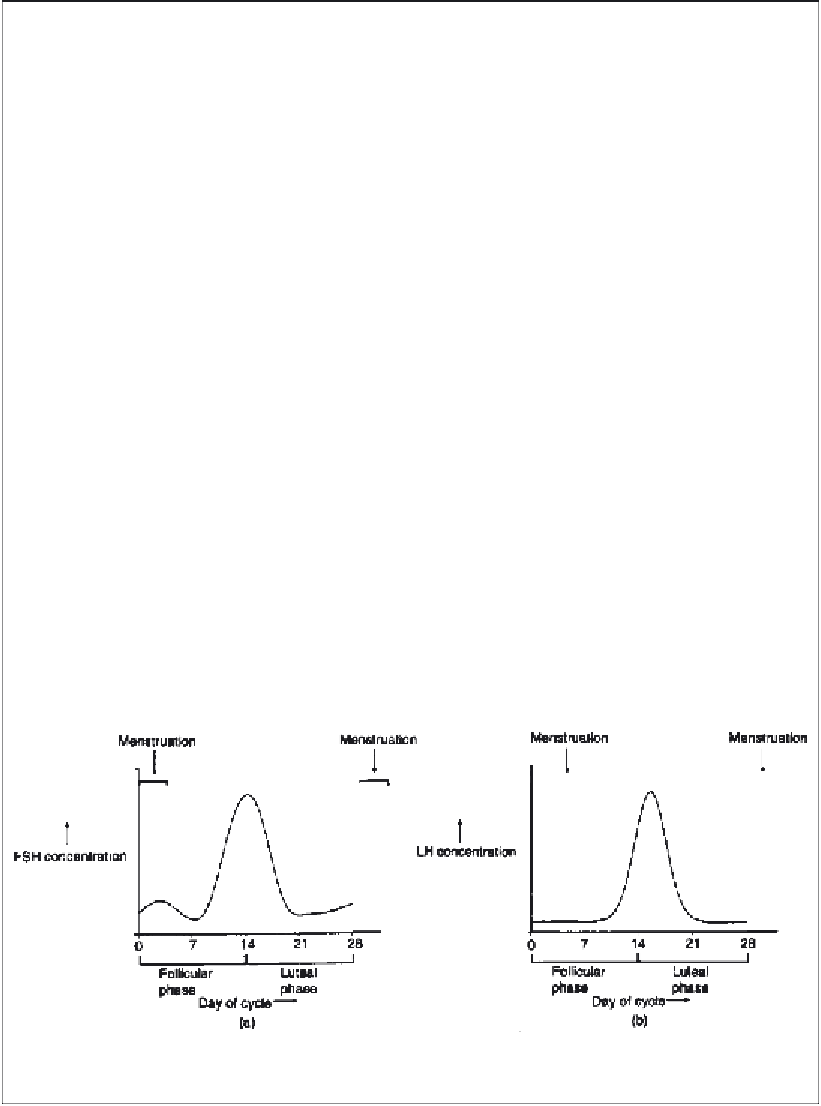
Figure 11.B1
Changes in plasma FSH (a) and LH (b) levels during the reproductive cycle of a healthy
human female
Search WWH ::

Custom Search