Biomedical Engineering Reference
In-Depth Information
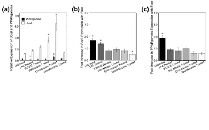
Fig. 5 Cytoskeletal tension signaling mediates MSC adipogenesis and flow-induced PPARc
expression. a Cytoskeletal dynamics affected both PPARc and Sox9 basal expressions. LPA
treatment that activates RhoA tension signaling decreased PPARc, while disrupting actin by
cytochalasin increased PPARc. Sox9 expression showed similar change under cytochalasin.
b Fluid flow-induced Sox9 expression is decreased by pharmacological agents that inhibit ROCK,
myosin II ATPase, and actin. c Fluid flow-induced PPARc expression was decreased under all
pharmacological agents, suggesting an intact cytoskeleton signaling may be needed for flow-
induced PPARc expression. Reprinted with permission from the Company of Biologists Ltd. [
2
]
pharmacological agents that inhibit cytoskeletal formation, flow-induced
upregulation in Sox9 and PPARc was attenuated (Fig.
5
). Combined, it was pro-
posed that RhoA-mediated cytoskeletal tension is a negative regulator of adipo-
genic differentiation of MSCs in static culture, whereas an intact, actin cytoskeletal
dynamics may be required for flow-induced gene expression even for PPARc.
It was recently reported that fluid flow applied to MSCs may produce similar
results of adipogenesis inhibition as with stretch and compression. MSCs were
stimulated by flow shear stresses using a multishear microfluidic device and it was
shown that fluid flow affects the regulation of yes-associated proteins (YAP) [
57
].
By increasing the magnitude of fluid shear stimulation, the expression of YAP was
increased leading to decreased adipogenesis, greater osteogenesis, and initiating
dedifferentiation for chondrocytes.
The other fluid flow studies for MSCs have mostly focused on the direction of
MSCs toward osteogenesis. These studies have hypothesized that biomimetic flow
conditions to which bone cells are exposed in vivo may help MSCs to commit
toward osteogenic phenotype. Bone cells in vivo are exposed to load-induced
matrix deformation and resultant interstitial fluid shear through lacunar-canalicular
microscale channels. Given that fluid flow stimulation stimulates the osteogenic
activity of mature bone cells and promotes healthy bone homeostasis, fluid flow
may also stimulate MSCs to differentiate into the osteogenic fate [
14
]. Recent
studies have shown that fluid flow enhances mineralized bone matrix deposition
and osteogenic gene expression in MSCs [
31
,
55
]. In these studies, key osteogenic
markers including AP activity, Runx2, osteopontin, BMP2, etc., were upregulated
in MSCs as a result of fluid flow stimulation. These suggest a potential of flow
shear as a vital trigger for MSC osteogenesis.
Search WWH ::

Custom Search