Biomedical Engineering Reference
In-Depth Information
A
Medial:
Lateral:
Medial > Lateral
Normal
BMI
P
Thickness
(mm)
4.4
M
L
Medial:
Lateral:
Medial > Lateral
BMI: High > Normal
0
High
BMI
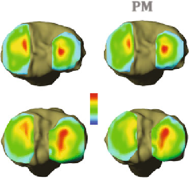
Fig. 2 Effect of body mass index (BMI) on diurnal changes in proximal tibial plateau cartilage
thickness as determined by 3T magnetic resonance images. Heat maps of cartilage thickness
measured in the morning (8:00 AM) and afternoon (4:00 PM) in representative normal and high
BMI individuals. Compressive diurnal cartilage strain was significantly greater in both the medial
and lateral plateaus in individuals with a high BMI ([25) compared to those with a normal BMI
(\25) [
42
]. Images kindly provided by Dr. Louis E. DeFrate
and under increased loading conditions, cartilage may up-regulate anabolic path-
ways to increase proteoglycan content and thickness [
36
]. Both local contact
anatomy and functional dynamic loading contribute to inter-individual differences
in cartilage thickness [
37
]. For example, in younger individuals (\35 years old),
femoral cartilage thickness is correlated with static and dynamic estimates of joint
stress levels, such as body weight 9 height and peak knee adduction moment,
respectively [
38
]. However, with obesity or increased age ([35 years old), this
association between joint stress load and femoral cartilage thickness is lost [
38
].
Thus, changes in how chondrocytes respond to mechanical stimulation due to
obesity and aging is expected to affect the relationship between joint loading and
tissue strain.
In vivo imaging techniques, such as magnetic resonance imaging (MRI), pro-
vide important insight into the effect of joint loading and disease status on articular
cartilage strain [
39
,
40
]. Recent studies have used MRI-based cartilage thickness
measurements in the morning and afternoon to examine diurnal strains in knee
articular cartilage (Fig.
2
). Diurnal strain measurements reflect local changes in the
cartilage mechanical environment resulting from accrued joint loading due to both
daily physical activity levels and gait dynamics [
41
]. In asymptomatic normal-
weight adults, articular cartilage undergoes a net compressive strain throughout the
day in the medial tibial plateau and the medial and lateral femoral condyles [
42
].
Age-, sex-, and daily step-matched overweight and mildly obese individuals
develop diurnal compressive cartilage strains in these same sites as well as in the
lateral tibial plateau (Fig.
2
). A higher body mass index (BMI) increased diurnal


Search WWH ::

Custom Search