Biology Reference
In-Depth Information
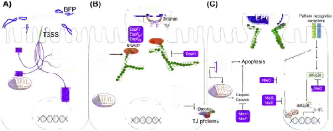
FIGURE 4.2
A model of EPEC attachment and host cell subversion: (A) Typical EPEC uses bundle-forming pili (BFPs) to attach to intestinal epithelial cells and
translocates effector proteins (purple) via a type 3 secretion system (T3SS). (B) Effector proteins serve as receptors (Tir) for intimin, subvert the host cell cytoskeleton
(EspF, EspF
u
, EspF
m
, MAP, EspT, EspM, EspV), interacting with host proteins such as N-WASP and the Arp2/3 complex to cause actin rearrangement and the forma-
tion of a cup-shaped pedestal, and disrupting barrier function (EspF, MAP, NleA, EspB) by inducing the redistribution of tight junction (TJ) proteins. (C) In addition
to altering the regulation of ion transport (not shown), EPEC effector proteins also modulate host survival responses; both promoting (EspF, EspG, EspH, MAP)
and antagonizing (NleF, NleH) apoptosis and inhibiting innate immune responses (NleB, NleC, NleD, NleE) activated by pathogen-associated molecular patterns.

Search WWH ::

Custom Search