Biology Reference
In-Depth Information
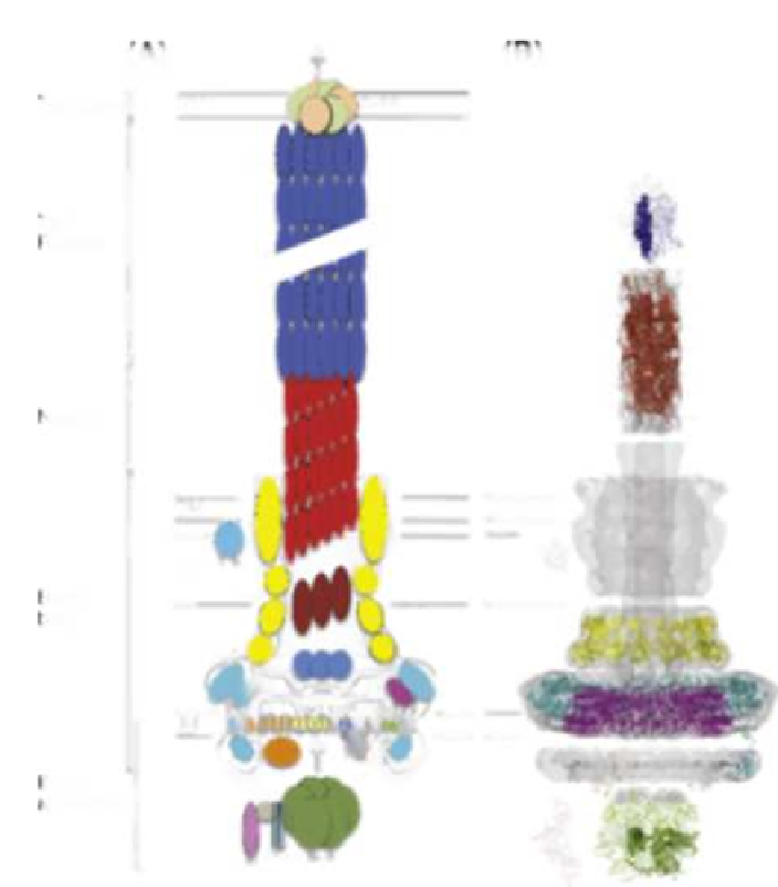
FIGURE 14.2
Structural organization of the T3SS injectisome. (A) Cartoon representation of the
various proteins constituting the injectisome, in the EPEC T3SS. The location of the injectisome
substructures (export apparatus, basal body, needle, filament, translocon) is indicated on the left.
IM - inner membrane, PG - peptidoglycan, OM - outer membrane, HM - host membrane. (B)
Known structures for individual injectisome subunits, in their putative localization relative to the
EM map. Structures are from a variety of species and systems (see the text for detail).
(
Marlovits et al., 2004
;
Sanowar et al., 2009
;
Spreter et al., 2009
;
Schraidt
et al., 2010
), EscJ is presumed to form the innermost ring component and,
given the lipoprotein signal peptide at the N-terminus (
Crepin et al., 2005
), is
localized to the inner membrane by a lipidation anchor (orthologs of EscJ in sev-
eral T3SSs also possess an additional transmembrane helix at the C-terminus)
(
Kimbrough and Miller, 2000
;
Schuch and Maurelli, 2001
). The EscJ struc-
ture consists of two independent small α/β domains of similar fold, joined by

Search WWH ::

Custom Search