Biomedical Engineering Reference
In-Depth Information
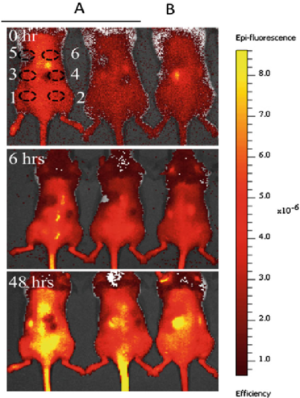
Fig. 4
Imaging of increased but delocalized inflammatory cell proliferation signals. Mice were
implanted with various inflammatory and non-inflammatory materials. ATX-Red was applied and
fluorescent images of the following time points: 0, 6 and 48h. First two mice from left were with
implants and the third mouse is without implants. Implants were as follows:
1
Porous glass implant,
2
Porous glass implant with dead bacteria,
3
Poly-L-lactic acid,
4
Magnesium,
5
Mock and
6
Titanium.
Dashed circles
represent the site of implantation
the dye injection at a site where there was no implant and which may correspond to
the location of the spleen (Fig.
4
, 48h, yellow spot on the left side of each animal).
However, there was a high background signal and the inflammatory signals were
delocalized, making it difficult to determine the response to individual implants.