Biomedical Engineering Reference
In-Depth Information
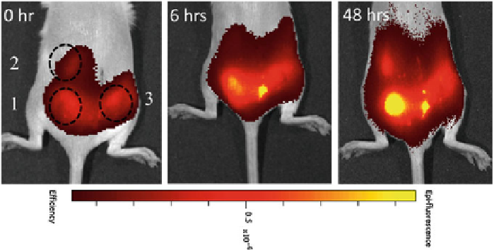
Fig. 2
Enhanced oxidizing potential in response to inflammatory implants visualized by a fluores-
cent probe. Biocompatible glass implants and glass implants coated with inflammation-inducing
heat inactivated
S. aureus
were subcutaneously implanted into Balb/c mice. Hydrocyanine solutions
were injected at the site of implantation as oxidation-sensitive dye precursor molecules. Fluores-
cence intensity was determined at 0, 6 and 48h after dye injection. The following implants were
used:
1
Glass bead coated with inactivated bacteria,
2
Glass implant,
3
Surgery without implant.
Dashed circles
represent the site of implantation
3 Results
3.1 Imaging of Inflammatory Oxygen Radicals
To visualize and compare inflammatory reactions over the time, biocompatible and
inflammatory material samples were implanted subcutaneously in mice. Reactive
oxygen products of neutrophil and macrophage cells were detected after subcu-
taneous administration of hydrocyanines. These reduced dyes become fluorescent
after an oxidation reaction with oxygen radicals. As highly inflammatory material
porous glass beads loaded with heat-inactivated bacteria were used. Fluorescence
intensity increased during first two days (Fig.
2
). The site with the bacteria appeared
brighter fluorescent when compared to bare glass implants. Therefore, oxidation sen-
sitive dyes could be used to differentiate inflammatory and biocompatible implants.
However, the response to a biocompatible implant and the wound healing response
showed similar fluorescence intensities and could not be differentiated by this method
(Fig.
2
).
3.2 Imaging Protease Activity in Inflammation
Proteases are secreted by immune cells and essential to allow migration through
the tissue. To image inflammatory protease activity in vivo, inflammatory and