Biomedical Engineering Reference
In-Depth Information
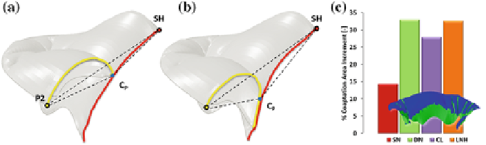
Fig. 4
Representation at peak-systole of the triangle of coaptation in pre-operative conditions (
a
)
and after neochordal implantation of a classic loop (
b
);
c
relative percentage CoA increment after
different NCIs
with the pre-operative model. The triangle of coaptation (as reported through dotted
lines in Fig.
4
a, b) is defined connecting the two points sited on the mitral annulus
(SH on the septal and P2 on the lateral mitral annulus) with the coaptation point
C
P
. In addition, all NCI simulations restored a comparable level of CoL (range:
6.2-7.2mm) with the largest values of CoL obtained when adopting multiple NCI.
As underlined in the histogram of relative CoA increment (Fig.
4
c), the use of
multiple neochordae, with respect to the
Pre-Op
model, progressively improved the
repair in terms of prolapse reduction and coaptation area recovery, passing from SN
to multiple NCIs, and determined a wider realignment of the free margin along the
P2 prolapsing region.
In all of the NCI models, a slight decrease in PMs reaction forces was noticed:
in Fig.
5
a the temporal curve of PMs reaction force is reported throughout the entire
duration of MV closure simulation, i.e. from initial valve closure to the final systolic
peak.
As regards chordal tension, in the prolapsing region, the tension decreased on both
marginal and basal native chordae, as starred in red and black color in the histogram
(Fig.
5
c) where chordal tension is reported at peak systole for all native chordae along
the posterior leaflet (the prolapsing region is located between themidline of P2 scallop
and the lateral P3 scallop) and for ePTFE sutures. As a consequence, tension load was
partially transferred from native chordae to artificial neochordae, this effect being
more emphasized when NCI with multiple neochordae was simulated.
In the
Pre-Op
model, a peak of MaximumPrincipal Stress was noticed on the free-
margin of the prolapsing scallop in proximity of a native chorda (Fig.
6
a) whereas
the prolapsing portion of the leaflet was partially unloaded.
In post-operative models, leaflet stresses along the non-prolapsing scallops were
substantially unchanged between pre- and post-operative analyses, regardless of the
employed technique. On the contrary, stresses on the prolapsing scallop markedly
changed: regardless of the simulated NCI technique, leaflet stresses were reduced
in the areas subtended by native chordae located in proximity of the prolapse and
increased in the repaired portion of the posterior leaflet since ePTFE sutures restored
mechanical tension along the prolapsing region of the leaflet (Fig.
3
). Moreover, as