Biomedical Engineering Reference
In-Depth Information
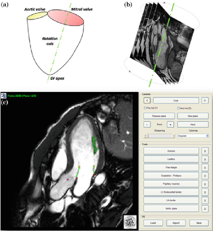
Calif) machine: cine-images were obtained through 18 long-axis planes, evenly
rotated along the left ventricle (LV) axis every 10degrees, with an isotropic
pixel
spacing
, stored in each DICOM image file as the distance between adjacent pixels,
of 1.25mmand a slice thickness of 8mm. Using a breath-holdingmodality, 30 cardiac
phases were recorded on each plane, with different temporal resolution according to
the characteristic R-R interval of each patient.
The use of a CMR rotational sequence of acquisition allowed to completely
acquire MV apparatus and dynamically assess its function through out an entire
cardiac cycle. The axis of rotation coincided with LV axis and it was defined as the
segment connecting LV apex and mitral valve centroid (as schematically reported
Fig. 1
CMR-acquisition protocol and offline segmentation:
a
positioning of LV long-axis of acqui-
sition;
b
derived rotational sequence of acquisition consisting of 18 long-axis planes;
c
view of the
segmentation panel with a tracking example of MV apparatus