Biology Reference
In-Depth Information
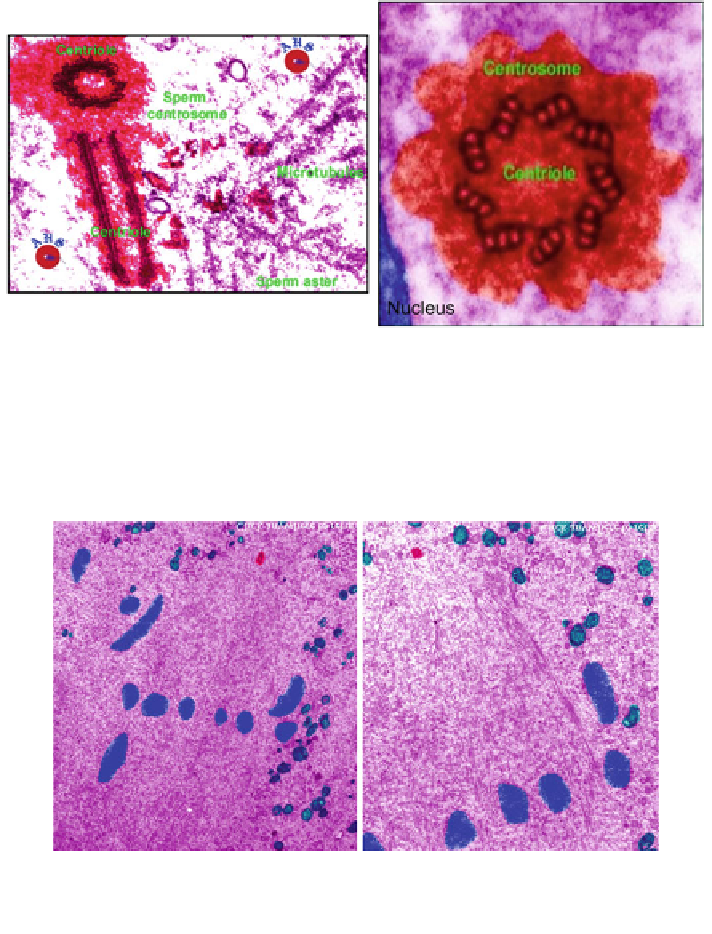
Fig. 5.19 Centrioles in monospermic embryos (TEM): Zygote centrioles associated with a
sperm aster soon after fertilization. It is now functional with duplicated centrioles and PCM (red)
nucleating MTs (left). A classic image of a juxta-nuclear centrosome in an eight-cell blastomere
(right). The centriole is located in the heart of the centrosome. The zygote centrosome is
replicated and perpetuated in embryos, fetal, and adult cells. Every cell has two centrioles.
Colored by computer x50,000, x170,000 (Sathananthan et al. 1966)
Fig. 5.20 Bipolar spindle at syngamy in a dispermic ovum showing centrioles at either pole
(red). Note chromosomes (blue) at the equator and few outside the spindle zone, composed of
MTs. Mitochondria are green. Computer coloured. x12,750 (Sathananthan and Edwards
1995
)
5.8 Centrioles in Embryos
Embryos cleave by repeated mitoses involving centrosomes. Descendants of the
sperm centrosome were found at every stage of preimplantation development and
were traced from fertilization through the first four cleavage cell cycles to the
morula and hatching blastocyst stages (Sathananthan et al.
1996
; Sathananthan