Biology Reference
In-Depth Information
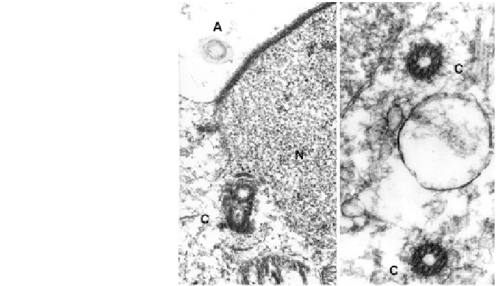
Fig. 5.11 Round spermatids
showing centrioles,
(C). They duplicate (left) and
migrate to the pole opposite
the acrosome, prior to
spermiogenesis (right)
A acrosome cap N nucleus.
x3,400, x85,000
5.6 The Maternal Centrosome
Unlike the sperm centrosome, the centrosome in the mature human oocyte is both
reduced and inactivated to become nonfunctional in the meiotically arrested oocyte
(Figs.
5.15
,
5.16
). The metaphase II spindle, at either pole, neither contains cen-
trioles nor dense, granulovesicular, centrosomal material that clearly nucleate MT in
mouse oocytes (Fig.
5.17
), which do contain a functional maternal centrosome.
Centrioles are generally absent in mammalian oocytes and are also not found in
mouse sperm (Sathananthan
1996
; Manandhar et al.
2000
). Although the mature
human oocyte has no visible centrosome, a functional centrosome with two typical
centrioles is found in fetal oogonia (Fig.
5.15
), which conforms to those of other
somatic cells (Sathananthan et al.
2000
,
2006
). These are, as usual, juxta-nuclear in
position, have PCM which nucleate MTs and seem to organize the oogonial cyto-
skeleton (a true centrosome). Therefore, reduction and loss of the maternal cen-
trosome has to occur either during oogenesis or in the final stages of oocyte
maturation when the first polar body (PB1) is abstricted, as in starfish oocytes
(Sluder and Rieder
1993
). We have examined several maturing oocytes by TEM and
have not yet located a centriole in PB1, nor in immature germinal vesicle oocytes. It
seems possible that the human follows the starfish pattern of maternal centrosomal
reduction since both follow Boveri's rule of paternal centrosomal dominance and
inheritance. The sequence of events of centriologenesis during spermiogenesis and
oogenesis are also available on the Web
www.sathembryoart.com