Biology Reference
In-Depth Information
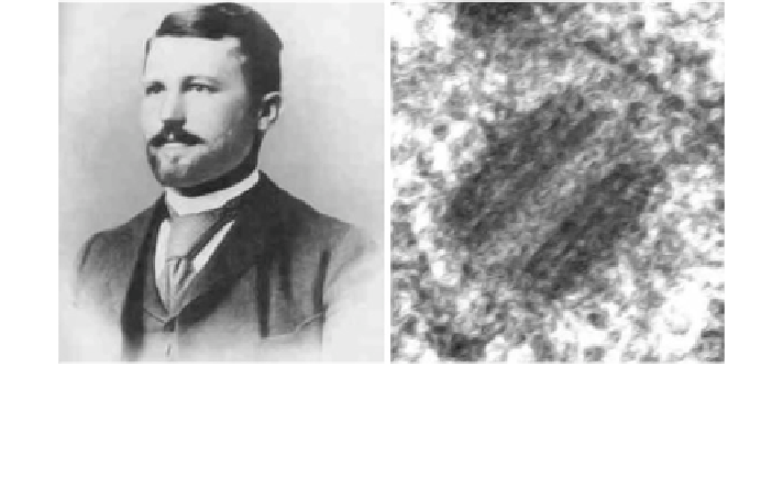
Fig. 5.1 Theodore Boveri (1887, 1901)—The father of the centrosome and his predictions,
based on his research on Ascaris and the sea urchin. Right Ascaris centrosome in a spermatocyte
TEM x200,000. Note its remarkable resemblance to the human centrosome (Sathananthan et al.
2006a
,
b
)
in cytokinesis during the cell cycle (Fig.
5.2
). The centrosomal cycle is closely
integrated with the chromosomal cycle in embryonic and somatic cells (Mazia
1987
). It plays a significant role in the cell cycle and cell division in most cells.
Like chromosomes, centrioles are self-replicating organelles which duplicate
during interphase, when they are located close to the nucleus. The process of egg
activation by sperm and initiation of embryonic cleavage was little understood,
until we discovered the important role of the centriole in humans in 1991(Satha-
nanthan et al.
1991
; Sathananthan
1991
). We first detected a centriole in a human
embryo in 1986 (Chen and Sathananthan
1986
) and this prompted us to investigate
this organelle in the following years. Pioneering work on centrosomes originated
in Australia in the early 1990s.
5.2 Ultrastructure
Transmission electron microscopy (TEM) is still the best way to study centrioles,
which are minute organelles (*2 lm in diameter), barrel-shaped, and presenting a
unique '9 ? 0' organization of microtubules (MTs) resembling a pin-wheel.
Centrioles are typically surrounded by pericentriolar material (PCM) that nucleates
MTs in somatic cells (Figs.
5.3
,
5.4
). The centriole and PCM complex (centro-
some) becomes functional in the oocyte only after fertilization, when the sperm
centrosome forms a sperm aster and the duplicated zygote centrosome forms the
zygote aster (Sathananthan et al.
1996
; Schatten
1994
; Simerly et al.
1995
).
Centrosomes are oftentimes indirectly localized using fluorescent microscopy