Biology Reference
In-Depth Information
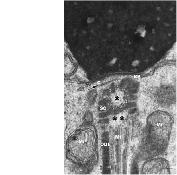
Fig. 2.3 Human sperm
connecting piece. The
proximal centriole (*),
sectioned at right angle, is
enclosed laterally by
segmented columns (SC) and
cranially by the capitulum
(arrow, C) which is lodged in
the implantation fossa at the
caudal pole of the sperm
head. A dense basal plate
(BP) lines the outer leaflet of
the nuclear envelope at the
implantation fossa. Distal
ends of SC are continuous
with outer dense fibers (ODF)
of the sperm axoneme.
Axonemal microtubules (Mt)
end cranially in a rarefied
area formerly occupied by the
distal centriole (**).
Mitochondria (Mi). Bar
represents 0.1 lm (Figure 3
was originally published by
Chemes et al. (
1999
) and
reproduced, modified from
the original, with permission
from the publisher)
migration-attachment of basal bodies-flagella will result in misalignments of the
tail and serious structural and functional sperm anomalies.
The growth of the sperm axoneme is accompanied by complex modifications in
the dense PCM. In its place, new proteins organize in nine longitudinal segmented
columns (SC) and the capitulum (C) of the connecting piece (Fig.
2.3
) (Fawcett
1981
). SC and C constitute a dense shield that lodges and encloses both centrioles.
The SC are nine cylindrical structures with periodic densities that fuse cranially to
form the capitulum, a curved plate-like disk that links connecting pieces to sperm
heads by its association to basal plates, dense structures that line the outer nuclear
membrane at the implantation fossa. At their caudal end each SC is continuous
with one of the nine outer dense fibers (ODF) that associate to peripheral
microtubular doublets of the growing axoneme. In many mammals, including
humans, the distal centriole vanishes after giving rise to the sperm axoneme,
leaving few remnants in mature spermatozoa.