Biology Reference
In-Depth Information
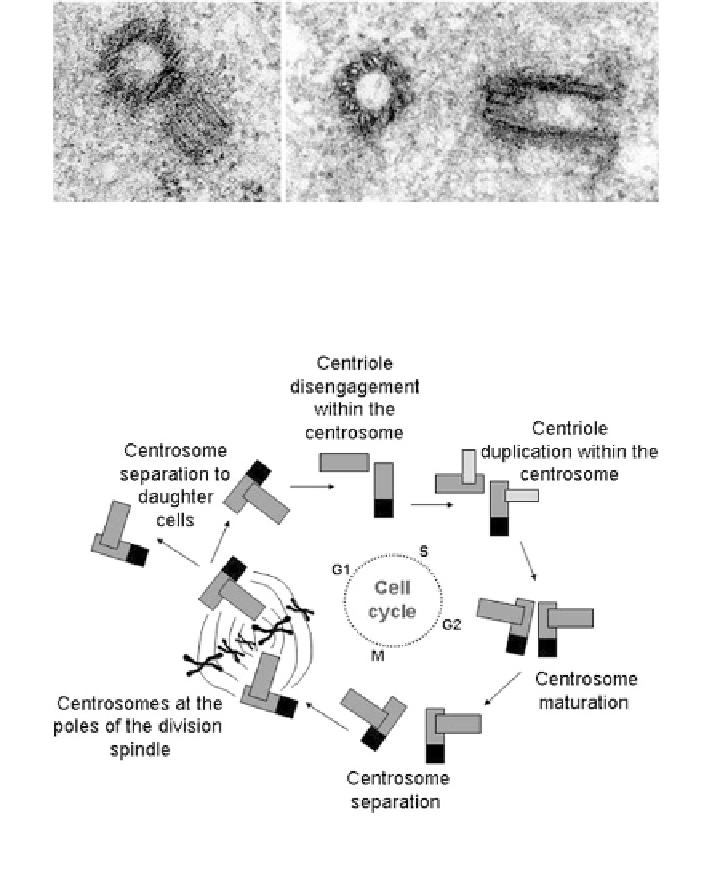
Fig. 20.1 Electron microscopy images of centrioles in Xenopus laevis oogonia. Left panel: pair
of closely apposed centrioles; Right panel: pair of separated centrioles. In both panels a
presumably mother centriole (on the left) is shown in cross section and a daughter centriole (on
the right) in longitudinal section. Courtesy of Malgorzata Kloc, Houston, TX
Fig. 20.2 Centrosome cycle in relation to the cell cycle progression. The mother centriole is
black and gray, daughter is gray
two are connected by intercentriolar links embedded in the PCM. Due to slow
growth and development of the daughter centriole the full reproduction cycle of
the centrosome covers two cell division cycles (Fig.
20.2
).
The structural cues have functional impact because the MT nucleation prefer-
entially occurs at the mother centriole and MTs are fixed to the appendages of the
mother. Also, the formation of a primary cilium belongs to exclusive capacities of
the mother centriole. Thus, the polarity-delivering centrosome is intrinsically
asymmetric assuring transmissibility of this cue to the cellular level.