Biology Reference
In-Depth Information
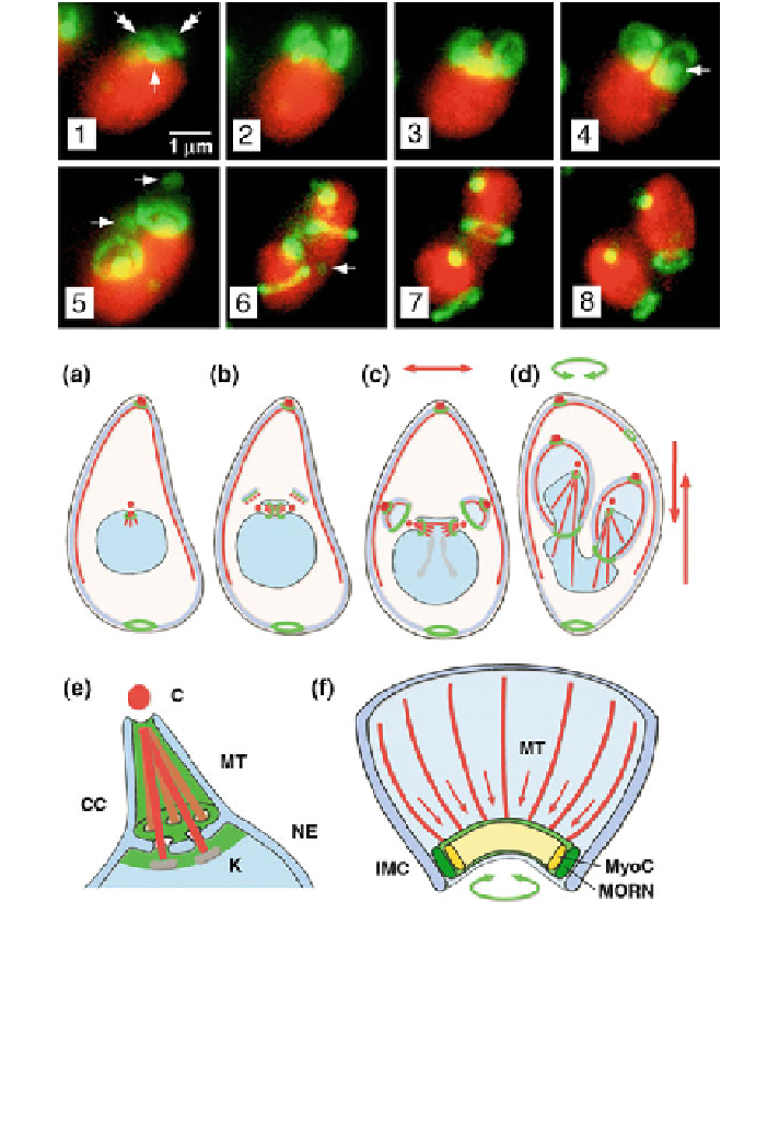
Fig. 19.5 Upper panel Fluorescent light microscopy images of a Toxoplasma strain expressing
TgMORN1 coupled to YFP (in green) throughout the parasite division process. The parasite
nucleus is stained in red. Arrows point to the centrocone (Modified from Gubbels et al.
2006
; with
permissions of The Company of Biologists). Lower panel Schematic model of the role played by
TgMORN1 in Toxoplasma division. Conoid (C), subpellicular microtubules (MT), centrocone
(CC) are highlighted in red, the nucleus in light blue and MORN1 in green. NE nuclear envelope,
K kinetochore, IMC inner membrane complex, MyoC myosin C. Red arrows indicate microtubule
driven movements and green arrows the constriction caused by MORN1 (Modified after Gubbels
et al.
2006
; with permissions of The Company of Biologists)
the TgMORN1-centrocone connection is observed throughout the Toxoplasma cell
cycle, although spindles have been found only during mitosis. Vaishnava et al.
(
2005
) described short spindles and centrocones through interphase in Sarcocystis