Biology Reference
In-Depth Information
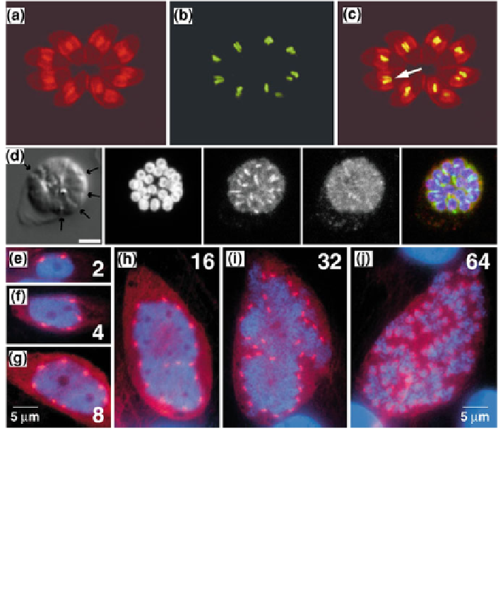
Fig. 19.2 Fluorescence light microscopy images of examples of the different division processes
in apicomplexa: endodyogeny (a-c), schizogony (d), and endopolygeny (e-j). a-c Toxoplasma
expressing an apicoplast molecule tagged with GFP (in green), and stained with an antibody
against the inner membrane complex (in red). The arrow in c points to the ''U'' shape of the
apicoplast during division (After Striepen et al. 2000; with permissions of The Rockefeller
University Press). d Plasmodium parasites (arrows) at the final stage of mitosis. The nuclei are
stained in blue, the MTOCs in red and the microtubules in green (After Gerald et al.
2011
; with
permissions of the American Society for Microbiology). e-j: Nuclear division and cytokinesis in
Sarcocystis. The a-tubulin is stained in red and the DNA in blue. Numbers indicate the number of
nuclei (After Vaishnava et al.
2005
; with permissions of The Company of Biologists)
must be well regulated in order to form new cells containing the correct set of
organelles and nuclear material. Endopolygeny is best characterized in Sarcocystis
neurona, a parasite of horses. During this process the DNA replication, nuclear
division, and cytokinesis processes are dissociated from one another, with five
cycles of DNA replication occurring prior to nuclear division, generating a 32N
nucleus (Vaishnava et al.
2005
). A final division generates 64 haploid daughter
cells. Curiously, the intranuclear spindle persists throughout the cell cycle. As will
be outlined below for other parasites, the apicoplast is associated with the
centrosomes and thus equally distributed to the daughter cells after replication.