Environmental Engineering Reference
In-Depth Information
1
B
C
2
e
e
3
h
A
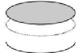
Figure 1.12
Diatom frustule struc-
ture - comparison of centric and
pennate diatoms.
Centric diatom
:
A. Separate views of epitheca (e:
1. Epivalve, 2. Mantle of epivalve,
3. Epicingulum) and hypotheca (h:
4. Hypocingulum, 5. Hypovalve). B.
Complete frustule - girdle or side
view (showing overlap of cingula). C.
Valve or face view of epivalve.
Pen-
nate diatom
: D. Complete frustule -
girdle view. E. Valve view, showing
apical or longitudinal axis (aa) and
transapical or transverse axis (ta).
h
4
5
aa
e
h
ta
ta
D
E
aa
under the light microscope (Fig. 1.13). The two pos-
sible valve views, of the top (epivalve) or bottom
(hypovalve) of the diatom, are not distinguishable in
many genera - but do differ in some cases (e.g.
Coc-
coneis
,
Achnanthes
). Axes of symmetry in pennate
diatoms are shown in Figs. 1.12 and 1.13.
overlying organic material to reveal frustule surface
structure.
The terminology of diatom morphology (see Glos-
sary, Chapter 4) includes various descriptors of
frustule markings - including eye-shaped structures
(ocelli), small pores (punctae) and fine lines (striae).
Illustrations of diatom surface markings are shown
diagrammatically in Figs. 1.13-1.14, and in various
figures and plates in Chapter 4. Although the bio-
logical significance of much of this surface detail
is obscure, the presence of one major surface struc-
ture - the raphe - is clearly associated with locomo-
tion. The secretion of mucus from this channel or
canal promotes movement on solid surfaces. In some
diatoms such as
Nitzschia
(Fig. 4.70a,b), the raphe
is elevated from the main diatom surface as a keel,
allowing more intimate contact between the raphe
and substrata. Such keeled diatoms are able to move
particularly well on fine sediments, and reach their
maximum abundance in the epipelon of pools and
slowly flowing streams (Lowe, 2003). A raphe is not
always present in pennate diatoms and is never seen
in centric diatoms.
Frustule markings
A wide range of surface markings can be seen on
the face (epivalve and hypovalve) of diatoms. These
have been recorded in considerable detail (Barber and
Haworth, 1981; Round
etal
., 1990; Wehr and Sheath,
2003) and form the basis for the classification and
identification of these organisms (see Chapter 4).
Clear visualisation of the frustule markings
requires the high resolution of either oil immersion
(light microscopy) or scanning electron microscopy,
and is normally carried out after the removal of
surface organic matter by chemical (acid diges-
tion) cleaning. Chemical fixation for scanning elec-
tron microscopy may also strip away some of the


Search WWH ::

Custom Search