Environmental Engineering Reference
In-Depth Information
50 μm
15 μm
50 μm
Figure 4.56
Ceratium.
Top: Two cells in fresh phy-
toplankton sample. Bottom: Scanning electron micro-
scope image showing typical dinoflagellate plated sur-
face (more clearly visible in Fig. 4.57). Cells have a clear
transverse furrow (arrow).
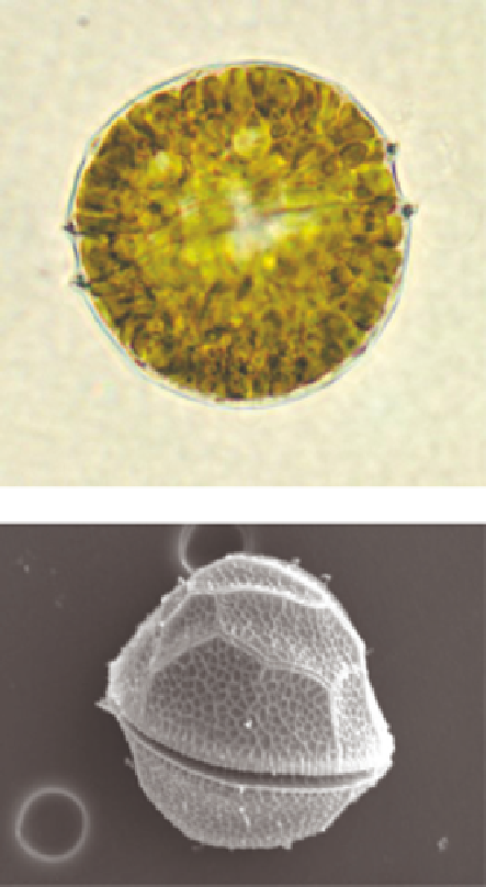
10 μm
Figure 4.57
Peridinium.
Top: Living cell containing
numerous brownish chloroplasts. Bottom: SEM image
showing surface outlines of thecal plates. Both images
show a clear transverse furrow (arrow).
(b) Cells elongate, cigar-, boat-, crescent-
shaped or a distorted version of any of
these. Decorations arranged bilaterally,
although this is not always obvious in cells
having a crescent or distorted shape. .
139
for positive identification. Since this facil-
ity will not be available to many users
of this text, features used in the key
will be generally limited to those visi-
ble using a good quality light microscope.
It is always best to observe both live
and cleaned diatom material (see Section
NB.
The structure of the diatom wall is
complex (see Chapter 1) and some fea-
tures may be difficult to see using light
microscopy alone. In many cases, scan-
ning electron microscopy may be required


Search WWH ::

Custom Search