Biomedical Engineering Reference
In-Depth Information
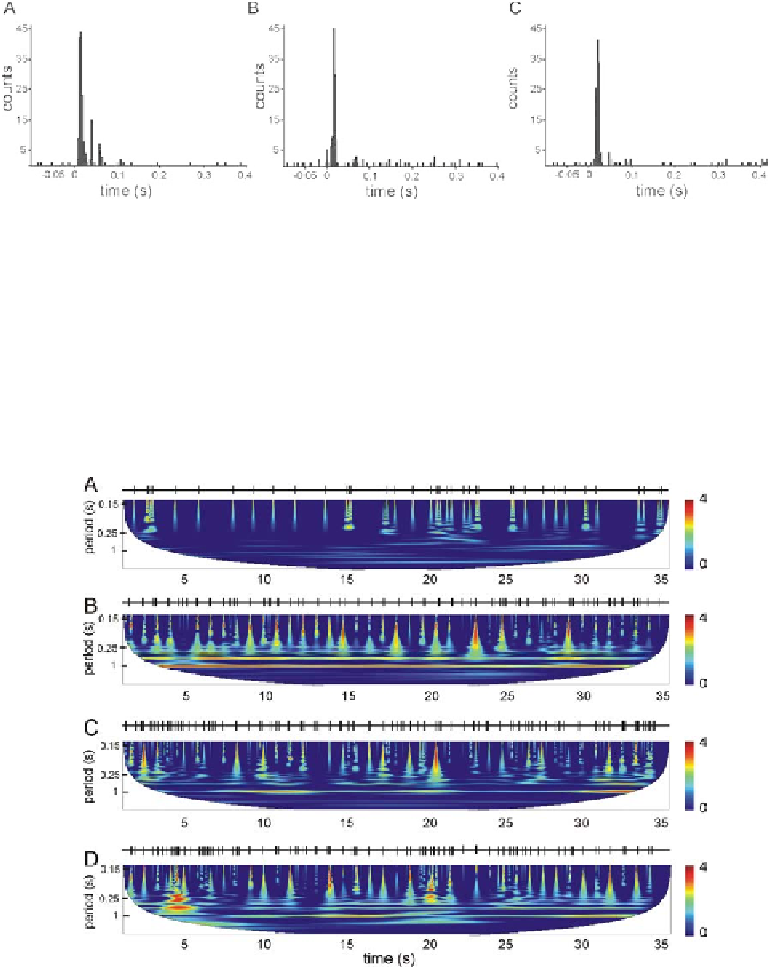
Figure 2. PSTHs of neural response to vibrissa deflection events in control conditions (A), and with
parallel distraction in ipsilateral (B) and contralateral (C) sides
Figure 3. Neural spike trains (the same as used in Figure. 2) and corresponding wavelet power spectra.
Color corresponds to the local spectral intensity. White area covers the cone of influence where the edge
effects cannot be ignored. (A) Spontaneous activity. (B) Response to periodic vibrissa deflection events (1
Hz rate) at control conditions. (C, D) Response to tactile stimulation with ipsilateral and contralateral
distraction, respectively. Tactile distraction reduces rhythmicity of the neural response to the vibrissa
deflection.
Search WWH ::

Custom Search