Biology Reference
In-Depth Information
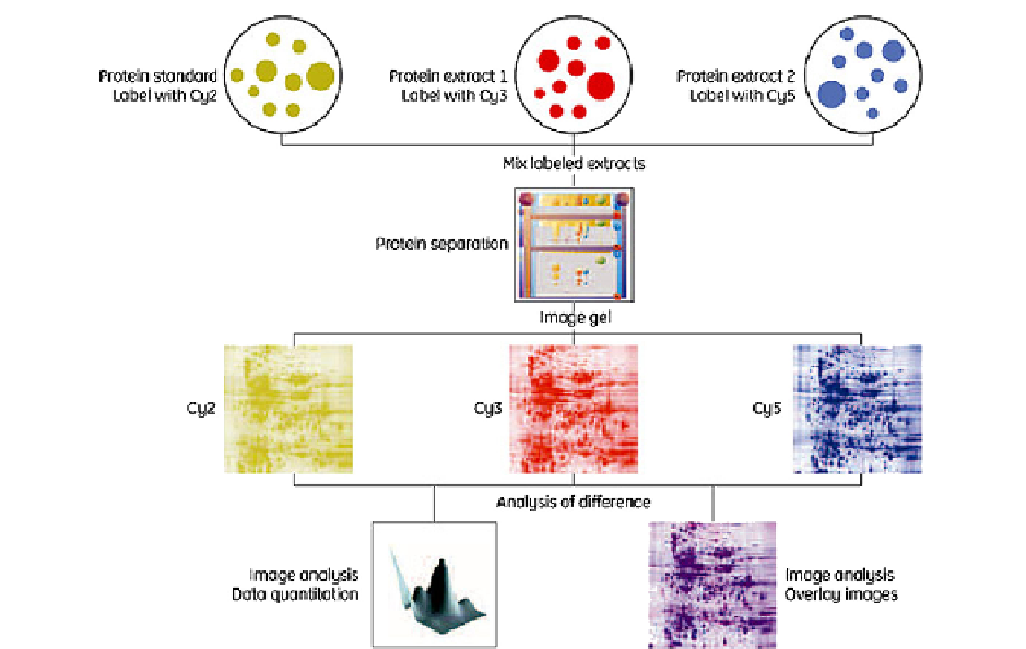
FIGURE 1
Schematic of 2D-DIGE procedure.
(Courtesy of Amersham Biosciences.)
APPLICATION OF 2D-DIGE TO
BIOMARKER DISCOVERY
Today, 2D-PAGE and 2D-DIGE play an
important role in disease biomarker discovery.
9
Petermann et al. discussed in a series of studies
published in the journal
Cancer
in 1948 the role
of plasma proteins in different types of cancer
using electrophoresis.
10
e
12
They concluded that
“
Two approaches are used in the search for
biomarkers: collected samples are analyzed (a)
individually or (b) pooled then analyzed. If blood
samples are used, the analysis is carried out pref-
erably by
none of the abnormalities found in these anal-
yses is characteristic of cancer in general or of
gastric cancer in particular.
first depleting the most abundant
proteins (HSA and IgG) using immunoaf
nity,
followed by labeling the proteins in the two
samples and internal standard by three different
cyanine dyes (Cy2, Cy3, and Cy5). The labeled
proteins are then spotted on the same gel plate
and separated. The intensity of the spots is
compared and the differentially expressed spots
digested into tryptic peptides. The peptides are
extracted from the gel and identi
In 1972, McIntire,
using gel electrophoresis, reported that serum
a
”
-fetoprotein is a biochemical marker for hepato-
cellular carcinoma.
13
The number of proteins
separated by electrophoresis before the develop-
ment of 2D gel electrophoresis was small and
included the high molecular weight proteins.
Iwaki et. al.
14
analyzed the proteome of urine
samples obtained from bladder cancer patients
and control subjects using 2D-PAGE. Three
proteins were identi
ed by high-
pressure liquid chromatography (HPLC) tandem
mass spectrometry andvalidatedbyWestern blot.
ed as novel tumor marker
Search WWH ::

Custom Search