Biomedical Engineering Reference
In-Depth Information
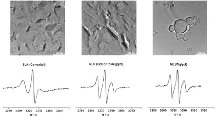
Fig. 4.12
Freeze fracture TEM images (
top
) and corresponding ESR spectra (
bottom
) of col-
loidal structures labelled with 0.025 % cholestane. (Composition: SLN (
left
)—glyceryl behenate,
NLC (
middle
)—glyceryl behenate/Miglyol, Nanoemulsion—Miglyol). Reprinted from J Control
Rel, Braem et al. (
2007
), with permission from Elsevier
signals arising from excitation of electrons in lipid nanoparticle dispersions. The
technique involves probing the molecular structure and organisation of supramo-
lecular systems and biomembranes. The ESR spectrum thus obtained provides
information about the sample microviscosity and the micropolarity.
It has been previously shown that storage-induced crystallization of lipid in the
lipid nanoparticles expels the probe out of the lipid phase into the outer aqueous
phase (Jores et al.
2003
). Accessibility of the lipophilic drugs to the aqueous phase
is indicated by the rapid loss of intensity of ESR signals. The time-scale of the
switch between the two phases can be exemplified by the ascorbic acid reduction
assay (Jores et al.
2003
). Reduction of the paramagnetic lipophilic drug to the ESR
silent hydroxylamine by the hydrophilic ascorbic acid is the underlying principle
behind this assay. ESR has also been used to study the interaction of SLNs with
membranes (Kristl et al.
2003
). Figure
4.12
shows TEM images obtained for vari-
ous colloidal structures and supporting evidence provided by ESR.
4.6 Conclusions
A wide variety of tools have been employed to study complex lipid nanoparticle car-
rier systems. The corroboration of results obtained from different techniques may
provide a better understanding of the structural and behavioral properties of these
systems. However, in vivo investigation of particle characteristics is often difficult.
The physicochemical properties of lipid nanoparticle dispersions are often dictated
Search WWH ::

Custom Search