Biomedical Engineering Reference
In-Depth Information
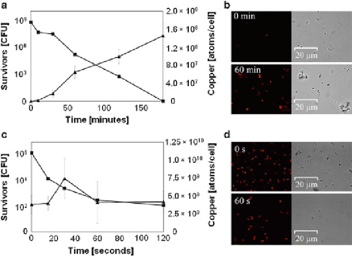
Fig. 6.5 Example of copper uptake on moist (a and b) and dry (c and d)by
E. coli
cells. Cells
were exposed to metallic copper surfaces for the indicated times, removed, washed, and plated on
solidified growth media. Survival was assessed by counting colony forming units (CFU) (squares
in a and c). In parallel, samples were mineralized and subjected to ICP-MS analysis to determine
cellular copper content (triangles in a and c) or were stained with the Cu(I)-specific fluorescent
dye Coppersensor-1 and subjected to fluorescence microscopy (b and d). Shown are averages
and standard deviations (
error bars
) from triplicate experiments (a and c) and representative
phase-contrast (
right
) and fluorescence (
left
) microscopy images (b and d)[
27
]
Cells experience a short sharp shock by contact with copper surfaces and a few
minutes are sufficient to completely inactivate all cells [
35
].
Under dry conditions, it was also noted that buffer composition or presence
of protectants influence the survival rate of bacteria. Cells applied with ROS
protectants (catalase, superoxide dismutase, manitol, etc.), chelators (EDTA), and
osmotic stress protectans (sucrose), increased survival on copper surfaces. This
topic will be a focus of the next section.
Survival Depends on Buffer Composition and Surface Corrosion
Under wet exposure, cells are suspended in a buffer that is in contact with the surface.
Composition of this buffer is important for copper ion release and, consequently,
killing efficacy differs in different buffer systems [
61
](Fig.
6.6
). Tris-buffer provokes
higher copper ion solubilization [
61
], hence cells become more sensitive to copper
Search WWH ::

Custom Search