Agriculture Reference
In-Depth Information
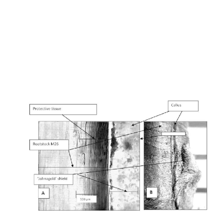
worth mentioning that a protective tissue, which consists of dead cells and is formed
in the place of wound near callus, remains without structural changes during the exis-
tence of a grafted plant and is not the spot where rootstock and a bud copulate.
The structure of a protective tissue is as shown in Figure 14.2. It consists of over
ten layers of elongated (up to 100 μm) fl at cells, which in this case are the remaining
elements of a leading rootstock system. Callus in the spot of copulation of a cultivar
bud and rootstock has typical yellow color, and it consists of parenchyma cells of vari-
ous size.
Analyzing Figures 14.1 and 14.2, when grafting is done between a bud shield and
rootstock section, leading system elements are formed only at a joint of callus forma-
tions. In fact, copulation of separate callus tissues occurs. Simultaneously, the process
of cell differentiation starts on some spots of callus tissue.
FIGURE 14.2
Longitudinal tangential cut (А) of the middle of the grafting place of cultivar
'Johnagold' on rootstock M.26 increased by 140 times; (B)—a general view of grafting.
The highest viability was typical for the cells, which were formed of cambium
and pericycle. Cross- and longitudinal sections of the grafting spot of vigorous apple
cultivar 'Gloster' on dwarf rootstock M9 after spring occurrence of a bud during inten-
sive growth and coloring of a leading system in methylene blue are shown in Figure
14.3. There (Figures 14.3(А) and 14.3(B)) one can see arteries of water with dissolved
nutrition elements in it, coming to a grafted bud. In a rootstock, elements of secondary
xylem and leading elements of the wood perform this function, whereas in a grafted
bud newly formed wedge-shaped leading elements do this. Where intensive callus is


Search WWH ::

Custom Search