Biology Reference
In-Depth Information
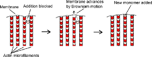
constantly with the object. The membrane is itself a 'small object' and, being flexible, its shape
will be continuously changing in response to the battering it receives from the molecules that
surround it. From time to time, therefore, the membrane will be moved forwards and the
space between it and the barbed end of a filament end and the membrane will open up; at
that moment a new actin monomer can join the filament (
Figure 5.13
). When the membrane
'tries' to move back, its way is now blocked by the elongated filament and its mean position
has therefore effectively been moved forward by the length of one actin unit. The process
then repeats. This basic mechanism remains valid whether the barbed end of the filament
actually contacts the membrane itself or contacts some cortical protein that in turn pushes
on the membrane.
The explanation above focused on the membrane alone; in reality, the terminal parts of the
filaments, too, will show random changes in length as they are compressed and stretched by
the molecules that hit them, and the ratchet mechanism is the result of both sets of random
motions.
Not all acrosomal processes develop by rapid polymerization of actin. In species such as
the horseshoe crab,
Limulus polyphemus
, the acrosomal actin is pre-assembled but is coiled up
in a tight spiral between the nucleus and the acrosome. In this cell, the actin is cross-linked by
two proteins: calmodulin and scruin which form a 1:1 complex. The complex changes confor-
mation depending on whether Ca
2
þ
is present in the cytoplasm. Receptors for the outer
surface of the egg trigger release of Ca
2
þ
in the sperm cytoplasm, and a change in conforma-
tion of the calmodulin/scruin complex. This relaxes the coiled actin bundle and it projects
forward to penetrate the egg's jelly coat.
56
One of the simplest types of protrusion made by ordinary mammalian cells is the small
microvillus. Small microvilli, which are shorter than 500 nm, are present in vast numbers
at the surface of most animal cells.
57
They are highly dynamic and short-lived structures,
each lasting only about 12 minutes. The dominant structure of a small microvillus is again
a bundle of cross-linked microfilaments that are packed in a hexagonal array. The microfila-
ments are orientated with their barbed ends towards the membrane and they grow from their
barbed ends, so that the already polymerized microfilaments are pushed towards the centre
of the cell. Normally, these filaments make connections with the cortical actin system and
therefore end up pushing against it rather than through it: the result is that the pointed
ends fail to move inwards and the barbed ends therefore have to move outwards, thus
creating a cell process (
Figure 5.14
).
FIGURE 5.13
The Brownian ratchet at the tip of the growing acrosomal process.