Biology Reference
In-Depth Information
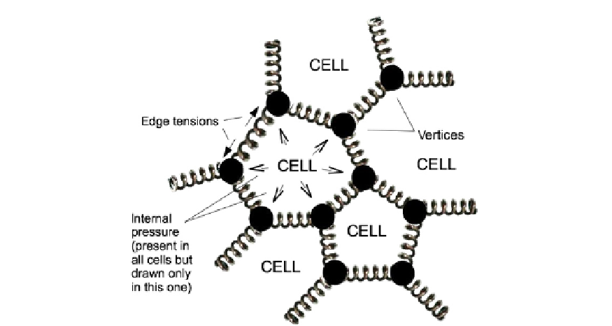
FIGURE 26.4
The structure of a typical agent-based model, in this case by Weliky and Oster (1990).
23
Each cell is
modelled as a source of pressure, bounded by straight 'springs' (tension-bearing elements) connected by vertices
(technically, a Voronoi polygon). Vertices move according to the balance of forces on them. Any 4-way vertices can
split (into two 3-way, connected by a new spring) if tension is high enough, or 3-way vertices can fuse to form
a 4-way one, eliminating the spring between them, if they approach within a threshold distance. The elements have
been drawn as springs for the sake of clarity, but this is not a literally physical model, existing instead only in
a computer. Also, there may be more vertices along the 'side' of a cell to represent more 'wiggles' of the
boundary
d
this diagram depicts a simple model with low numbers of vertices, for clarity.
plane: volume in a three-dimensional model) and its 'ideal' area, specified as a parameter of
the model. Cells that are squeezed therefore push back, while ones that are stretched out pull.
The probability and direction of a vertex moving is determined by the net pressure that acts
on it (each vertex will be acted on by at least two cells, many by three), by the angle of the
vertex (sharp angles encourage movement in most models, a feature that encourages protru-
sions from the ends of elongated cells) and by the 'stretch' in the spring. Diffusible factors
originating within the plane and from without can modulate the stiffness of springs in
a concentration-dependent manner.
Illustrating how computer models can be effective without running them is difficult, but
there are some examples, the results of which can, with hindsight, be understood without
a computer. An early application of agent-based models to study epithelial development
was to resolve a paradox during epiboly in
Fundulus heteroclitus.
16
Epiboly is a cell movement
in which a cap of about 3000 cells forming an epithelial monolayer on the top of the animal
pole of the egg embryo extends vegetally until it engulfs the whole thing (
Figure 26.5
a). The
epithelium is pulled by waves of contraction in the underlying yolk syncytial layer. The
spherical shape of the egg means that the circumference of the 'bottom' end of the cap
must expand continuously until it reaches the equator, and only then can it contract. The
paradox was that observation of embryos showed that the number of cells around the
bottom of the epithelial cap keeps decreasing during epiboly, when the circumference is