Biology Reference
In-Depth Information
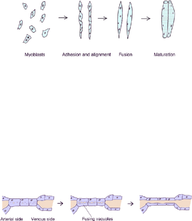
FIGURE 4.4
A schematic view of the cell alignment and fusion involved in the development of vertebrate
skeletal muscle.
they are induced, by signalling molecules such as Wnts and Shh, to form muscle they
multiply and align with each other to form long chains of cells. The cells within each chain
then fuse to produce a single multinucleate cell, the myotube, which matures into a long,
thin, muscle fibre (
Figure 4.4
).
12
An essentially similar process takes place in the formation
of somatic muscles in insects such as Drosophila melanogaster.
13
Cell fusion can also take place between one part of a cell and another part of the same cell.
This type of cell morphogenesis is used to create tubes (Chapter 19).
CELL CAVITATION
The finest blood capillaries of the body are so narrow that their lumens run through indi-
vidual cells rather than through tubes formed by multiple cells. The formation of lumens
begins when the cells concerned form large intracellular vacuoles by pinocytosis (
Figure 4.5
).
These vacuoles coalesce with each other and with the plasma membrane so that a hollow
lumen is formed,
14,15
and the cavitation is coordinated between neighbouring cells so that
their lumens join up to form a very fine blood vessel.
FIGURE 4.5
Cavitation of endothelial cells by fusion of vacuoles to create fine vessel lumens.
CHANGES IN CELL SHAPE CAN DIRECTLY DRIVE
MORPHOGENESIS OF TISSUES
All morphogenesis depends to some extent on changes of cell shape, but in some cases the
change in shape of a large-scale structure is driven directly by a change in the shape of the
cells that comprise it. These cases underline the more general importance of understanding
the basis of cell shape, if the shapes of organisms are to be understood.