Biology Reference
In-Depth Information
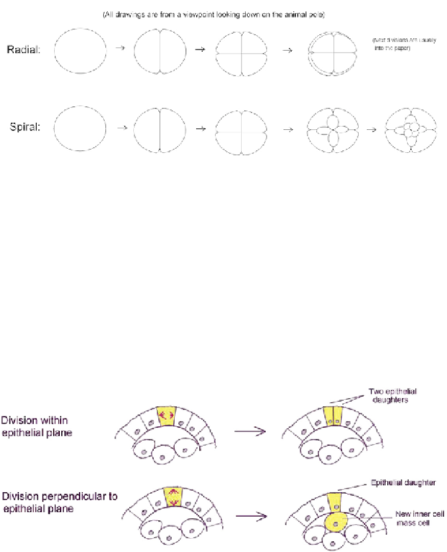
FIGURE 23.1
The two most common modes of cleavage in animal embryos. In radial cleavage, mitoses are
arranged at right angles to each other in the meridian or transverse planes of the embryo; they may produce equally
sized daughters, as shown here, or unequal ones. In spiral cleavage, mitoses proceed at an angle to the meridian and
transverse planes, and daughter cells are placed in the 'valleys' between other cells.
but they concentrate more on the energetics of the final cell shape than on the earlier placement
of the spindle that is, in most systems, the determinant of the cleavage plane (see below).
The models might therefore be viewed with some scepticism. It is possible
d
indeed
probable
d
that cleaving embryos use the same mechanisms used in later mitoses, as described
below.
The orientation of mitotic cleavage planes in epithelial tissues can have a profound effect
on morphogenesis (the general structure of epithelial tissues is discussed further in
Chapter 15). An example of this is seen in the first epithelium formed by a mouse embryo.
The cells of this epithelium, the trophoblast, can divide either in the plane of the epithelium
to produce two equal epithelial daughter cells or divide perpendicularly so that one daughter
remains within the plane of the epithelium while the other is driven in towards the centre of
FIGURE 23.2
Orientation of mitoses in the mouse trophoblast determines whether both daughters of a division
remain in the trophoblast or whether one joins the inner cell mass, which will give rise to the 'embryo proper'. This
diagram is based on the data in Pedersen et al.
5