Biology Reference
In-Depth Information
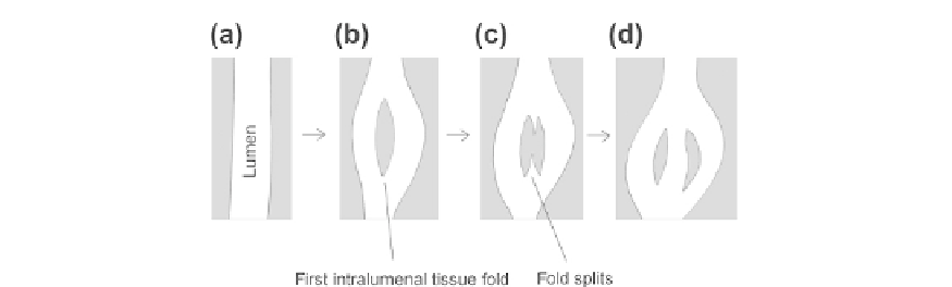
FIGURE 20.16
The splitting of an intralumenal tissue fold to create a new passage for blood flow. Again, the
lumen is white and the tissue grey.
One they have formed, the tissue barriers that divide a lumen can themselves split so that
blood flows through them, thus increasing the surface area for substance exchange even more
(
Figure 20.16
).
The cellular mechanisms of intussusceptive branching are not yet understood in detail, but
by drawing analogies with the processes of epithelial folding and fusion, it is possible to
make educated guesses that can be tested by experiment. The general decision to undergo
branching at all is probably regulated by the tissues that the blood system is there to serve.
Tissues that require a better blood supply, for example those that are anoxic, secrete factors
such as VEGF, FGF2 and angiopoietins that encourage both sprouting and intussusceptive
branching.
85,93
e
97
This will presumably create a general stimulus for branching activity but
would be unlikely to specify precisely where around the edge of a vessel an endothelial
fold/pillar should form. The positions of folds and pillars is probably determined at least
in part by the conditions of blood flowwithin the vessel, with the flow conditions either spec-
ifying the line of emergence of a fold or determining which of many random attempts to
make a fold can be stable enough to succeed.
The idea that blood flow can specify the location of a morphological event is not as far-
fetched as it may seem and there are several ways in which this might happen. Endothelial
cells are known to have a number of mechanisms by which they can sense the pressure and
shear of the fluid that flows across them and alter their gene expression accordingly.
97,98
It is
therefore possible that local measurements of flow act as inputs to the morphogenetic effec-
tors of folding (see below) and at least bias the probability of folds being initiated at particular
points. The pressure exerted by a moving fluid decreases as speed of movement increases,
)
and this could, in principle, also induce infolding by a direct sucking in of the walls which
could be stabilized by positive feedback (for example, cell bending driven by the fluid initi-
ating cytoskeletal changes that extend the fold).
The cellular mechanisms of fold formation have not been investigated in detail, but the
obvious analogy with epithelial invagination and evagination makes it likely that it is driven
)
This is the Bernoulli effect: it can be demonstrated very simply in the lab by hanging two ping-pong balls
on long strings so that they are next to each other, separated by a few centimetres. If a student blows between
the balls, to create air whose velocity is greater than that in the rest of the room, the balls draw
together
.