Biology Reference
In-Depth Information
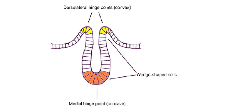
FIGURE 18.10
The neural plate folds mainly at hinge points, drawn here from a micrograph of avian neuru-
lation.
23
Note that the wedge-shaped cells marked in orange and yellow have opposite directions of wedging with
respect to their apico-basal polarity. For clarity, this diagram exaggerates the size of cells and in reality there are
many more of them, densely packed but still in a single layer.
involved, though, show that cytochalasins do not prevent the acquisition of a wedge shape at
the hinge points but rather interfere only with later development.
28
In mammals, formation
of the cranial neural tube is inhibited by cytochalasin D, but formation of the more posterior
neural tube is apparently unaffected by the drug.
29
This unexpected variation probably
warns that actin-based contraction cannot be the only mechanism for wedge formation.
The convex hingepoint cells of the neural tube, yellow in
Figure 18.10
, have to contract at
their basal rather than apical ends. In mice, Shroom is expressed under the regulation of the
secreted signalling molecule Sonic Hedgehog (Shh) and in shh
/
mutant embryos the
medial, concave hinge point fails to form, but the neural tube still forms and it closes under
the action of the dorsolateral, convex hinge points alone.
30
This strongly suggests that basal
constriction by the dorsolateral hinge cells is an active process rather than being a passive
consequence of bending by the medial hinge point. Observations made in cultured renal
epithelial cells show that targeting constitutively active RhoA to the basal sides of cells can
cause basal constriction analogous to the apical constriction seen when RhoA is targeted to
the apex.
5
This suggests one possible mechanism by which a convex hinge point may
form although, for reasons analogous to those depicted for apical construction in
Figure 18.11
,
there must be some additional mechanism that prevents the apex also becoming thinner,
allowing the cells undergoing basal construction to become columnar instead of doing the
work of bending tissues. Later in neural tube development, the neural tube itself undergoes
a crease-like invagination at the boundary between the midbrain and the hindbrain. The
formation of this crease is critical to subsequent morphogenesis of the brain and, because
the outer surface of the neural tube is the basal one, invagination involves basal constriction
at the centre of the crease.
31
In the fish Brachydanio rerio, the process begins by a local apico-
basal shortening of cells (a sort of 'opposite' to placode formation) and then proceeds by basal