Biology Reference
In-Depth Information
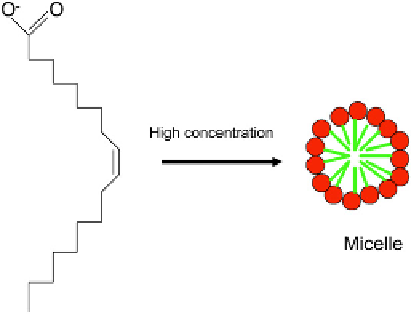
FIGURE 3.6
The formation of micelles by sodium oleate at high concentration.
lecithin (
phosphatidylcholine), were in constant motion and tumult for many minutes and,
indeed, when I was obtaining this image,
)
colleagues glancing at the VDU assumed that they
were seeing a complex living system.
The dynamism of vesicles has provided the basis of what is so far the most advanced
attempt to construct a self-reproducing 'cell' from non-living components. The starting
material is not a phospholipid but a smaller amphipathic molecule, sodium oleate
(
Figure 3.6
). At high concentrations, sodium oleate forms micelles in water. If a suspension
of such micelles is injected into a large volume of pH 8.8 buffer, the oleate rearranges to
form vesicles. The kinetics of this rearrangement show a sigmoidal curve
8
(
Figure 3.7
).
This curve, with an initial lag followed by a marked acceleration of vesicle formation once
some vesicles had already formed, indicates an autocatalytic process in which the presence
of existing vesicles encourages the conversion of micelles to vesicles. Furthermore, if pre-
existing vesicles are added to the system very early in its 'development', the lag phase is
markedly reduced and fast conversion of micelles to vesicles takes off immediately. Vesicles
therefore catalyse their own growth by 'feeding on' the micellar stocks of oleate (
Figure 3.8
).
The vesicles of this system are capable of more than simple growth. However, they can
also reproduce. The first evidence for this was the observation that, if vesicles of 100 nm
diameter are used to 'seed' the solution, the result is a larger number of vesicles of about
100 nm rather than the simple enlargement of 'seed vesicles'. This suggests that the vesicles
must be using newmaterial to produce further vesicles of their own average size, perhaps by
a cycle of growth and fission. Visual evidence suggestive of fission comes from cryo-electron
microscopy, which reveals the presence of paired vesicles.
8
Biochemical evidence comes from
experiments in which the 'seed' vesicles are tagged with ferritin, an electron-opaque marker.
9
¼
)
Dry a drop of lecithin, dissolved in chloroform, on a microscope slide. When it is fully dry, place a drop of
water so it touches the edge of the lecithin smear, and observe the interface by phase contrast. Interesting
morphologies will result in minutes: this can make an interesting lecture theatre demonstration if a projec-
tion microscope is available.