Biology Reference
In-Depth Information
The idealized geometry illustrated in the diagram above does not do justice to the complete
model, but detailed computer simulations have been carried out and they produce biologically
sensible results.
14
In simulations of mixed cells, those with higher interfacial tensions envelop
those with lower interfacial tensions. Reconciling this result with observations on the behav-
iour of cells expressing different amounts of Cadherin 2 would require higher levels of
cadherin adhesion to correlate with lower interfacial tensions. This may be counter-intuitive,
considering the close associations between adhesions and the contractile cytoskeleton, but is
not impossible given the many ways in which adhesion complexes can initiate signals that
modulate myosin activity. Unfortunately, no measurements on either interfacial tensions or
absolute adhesiveness, which would settle the matter, have yet been made.
Condensation of limb mesenchyme, driven as it is by Cadherin 2, probably also requires
some other mechanisms described in this chapter; for example, the interstitial matrix has to
be cleared out somehow, before new matrix of the cartilage is laid down. The patterning
of the limb, which sets up positional signals to ensure that condensation takes place at appro-
priate sites and times, is outside the scope of this topic (which restricts its focus to morpho-
genesis itself) but good reviews can be found elsewhere.
16,17
Adhesion-driven coalescence may involve more than one cell type, as occurs in the devel-
oping gonads of
D. melanogaster
. The cells of the gonads are of two broad types, somatic cells,
which develop from mesoderm local to parasegments 10
12, and germ cells that reach the
somatic cells after a long migration through the embryo (the migration is described in
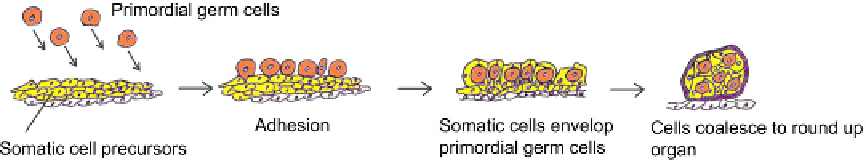
Chapter 12). During normal development, primordial germ cells arrive at and adhere to
the presumptive gonad when it is still quite an elongated structure. The somatic cells then
extend around the primordial gem cells and condense together to make a more rounded
and compact organ
18,19
(
Figure 14.5
).
Like the mesenchyme of tetrapod limbs, the coalescence of cells in the gonad depends on
homophilic adhesion molecules of the cadherin family, in this case the E-cadherin homo-
logue, DE-cadherin. Somatic gonad cell precursors and primordial germ cells both express
this molecule. In mutant embryos that lack DE-cadherin function, the primordial germ cells
fail to become ensheathed by the somatic cells and the presumptive gonad fails to round up
and remains in its elongated state.
19,20
The cells also express the transmembrane protein Fear
of Intimacy.
20
It does not seem to be needed by the germ cells, but if somatic cells lack it then
their levels of DE-cadherin are abnormally low and envelopment and rounding again fail.
The
D. melanogaster
gonad contains an important remaining mystery; since both primor-
dial germ cells and somatic cells express DE-cadherin, why do the germ cells not bind
e
FIGURE 14.5
Schematic diagram of gonad formation in D. melanogaster, based on Jenkins
et al.
19
In reality
there are more cells than depicted here (about 30 somatic cells per gonad).