Biology Reference
In-Depth Information
Drosophila melanogaster
, Fasciclins I, II, III and IVare expressed on the cell surfaces of different
axon bundles.
35
e
38
Fasciclin I, for example, is expressed initially in all commissural (midline-
crossing) axon bundles and later becomes restricted to a subset of them.
39
Disruption of fas-
ciclin expression or function disrupts fasciculation of axons,
40,41
while forcing abnormally
high levels of fasciclin expression prevents defasciculation where the axons ought to leave
the fibre bundle to fan out to their targets
42
. Only fibres using the particular fasciclin that
is subject to experimental manipulation are affected. (Fasciclins have other functions in later
neural development; for example, synaptogenesis, as well).
43
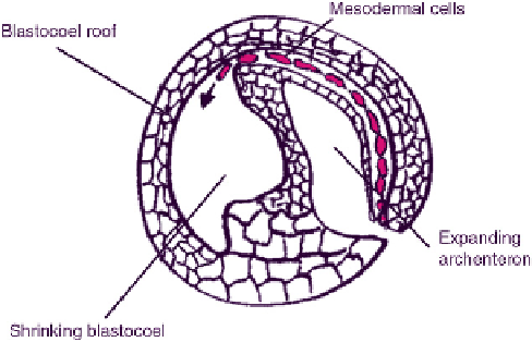
GUIDANCE OF CELLS BY ALIGNED FIBRES
During amphibian gastrulation, prospective mesodermal cells from the region of the blas-
topore migrate along the underside of the roof of the blastocoel towards the animal pole
(
Figure 11.7
). Shortly before this takes place, in both
Pleurodeles waltlii
and
Amystoma mexica-
num
, the roof of the blastocoel expresses high concentrations of the extracellular matrix
molecule, fibronectin.
44,45
The fibronectin is important for mesodermal migration, which fails
to take place when the embryo is injected with function-blocking anti-fibronectin anti-
bodies.
46
Antibodies that block the function of fibronectin-binding integrins have a similar
effect.
47
The blastocoel roof can be dissected from an embryo, cultured inside-down for
a while and then removed from its culture dish; what remains on the culture dish is an
acellular, fibronectin-rich matrix that was secreted by roof cells and is now 'blotted' on to
the dish. Remarkably, when mesodermal cells are placed on this matrix, they migrate
towards the part that underlay the animal pole whatever their original orientation
48
. The
matrix is therefore a sufficient directional cue. There is, however, no evidence for a large-scale
adhesive gradient in this matrix and cells seem to stick to all of its regions equally.
48
Electron microscopy reveals a potential directional cue in the micro-structure of fibro-
nectin in both living embryos and 'blotted' matrices in culture. Its molecules are aligned to
FIGURE 11.7
Migration of mesoderm cells under the roof of the blastoderm during amphibian gastrulation.
The figure depicts an embryo of
Xenopus laevis
part way through gastrulation
d
the other embryos mentioned in this
section differ in detail but not in the essentials of migration.