Biology Reference
In-Depth Information
in an earlier study that compared a commercial density phantom to rice as a soft-tissue equiv-
alent (
Cohen and Rushton, 1995
).
The box was positioned on the table so that the femur (right or left) was also in the approx-
imate anatomical position (about two-thirds down the table, mimicking a patient lying on the
table). In this way, the machine interprets a complete individual. Consequently, we could use
the standard DEXA software. The arm of the machine was brought to a level just superior to
midshaft. The areas of interest were manually selected on the computer by moving the rect-
angular field of view over the femoral neck. Two triangular fields of view were placed over
the greater and lesser trochanters. The standard DEXA measurements of bone mineral
density (BMD) (g/cm
2
) were calculated automatically for four anatomical areas: (1) the
femoral neck, (2) Ward's triangle, (3) the greater trochanter, and (4) the proximal shaft, in
addition to the calculation of total BMD.
CT Scanning
To facilitate CT data acquisition I worked with another anthropologist and biomedical
engineer to develop a system that permitted rapid data collection. Six identical sets of two
boxes (one 125 cm
5 cm) were built from
foam core board. Each set of two boxes was large enough to hold all the skeletal elements
32 cm
3 cm and the other 150 cm
22 cm
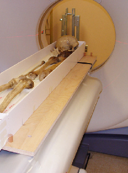
FIGURE 14.7
CT scanning boxes to standardize scanning positions.