Biology Reference
In-Depth Information
estimate age-at-death.
Stout and Stanley (1991)
tested both secondary osteon counts and
percent osteonal bone and found counts to be more accurate in predicting age estimates.
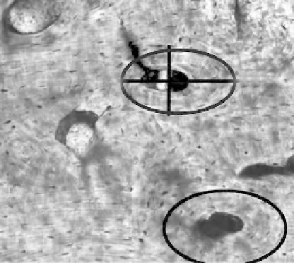
Secondary osteons are measured in several ways. Mean osteonal area, perimeter, and
maximum and minimum diameters have been used to predict age-at-death estimates
(
Thompson, 1979; Watanabe et al., 1998; Pfeiffer et al., 2006
). The Haversian canal of every
secondary osteon is similarly quantified utilizing average canal area, perimeter, and diame-
ters to estimate age (
Thompson, 1979; Thompson and Gunness-Hey, 1981; Watanabe et al.,
1998
). Research suggests that during aging, these variable measurements decrease in
response to increased bone remodeling and the addition of more secondary osteonal bone
(
Thompson, 1979; Thompson and Gunness-Hey, 1981; Watanabe et al., 1998; Pfeiffer et al.,
2006
). See
Figure 13.5
.
Additionally, researchers have looked at primary osteonal counts and fractional volume
of primary osteons as well as percent lamellar bone (
Kerley, 1965
; Ahlqvist and Damsten,
1969). The fractional volume refers to the percentage or ratio of bone comprised of primary
osteons versus secondary (or remodeled) osteonal bone. As age increases, the number of
primary osteons as well as the percentage of lamellar bone decrease and are replaced by
secondary osteons.
Other variables considered to estimate age-at-death include average cortical thicknesses
(
Thompson, 1979
; Thompson and Gunness-Hey, 1989), secondary osteon circularity (
Ortner,
1975; Britz et al., 2009
), and number of secondary osteonal fragments (
Kerley, 1965; Clarke,
1987; Ericksen, 1991; Cool et al., 1995
).
The reliability of these age estimators is dependent upon the individual bone (as
mentioned in the upcoming paragraph) and on methodology. Through decades of research,
the technique developed by
Kerley (1965)
and later fine-tuned (
Kerley and Ubelaker, 1978
)
has produced the most reliable age-at-death estimations. Kerley considered the following
variables: number of whole osteons, number of fragmentary osteons, percentage circumfer-
ential lamellar bone, and number of non-Haversian canals.
Stout and Stanley (1991)
found
FIGURE 13.5
Example of measurements taken by imaging software.