Biology Reference
In-Depth Information
at different rates by osteoblastic or osteoclastic activity on specific areas of bone. For example,
for long bones, osteoblasts increase the diameter of the shaft by adding bone to the periosteal
surface while osteoclasts remove bone from the endosteal surface (
Frost, 1964; Lacroix, 1971;
Sharpe, 1979; Martin and Burr, 1989
).
Prior to bone modeling, bone tissue is comprised of loosely organized woven bone.
Woven bone is immature bone with randomly oriented collagen fibers. It is temporary and
eventually replaced by lamellar bone. It generally disappears when children are very young
(
Martin and Burr, 1989
) and is usually not found in adults unless in response to pathology or
trauma. Woven bone can be rapidly produced to provide temporary mechanical strength
during skeletal repair. It is unique in that it is the only type that can be deposited de novo,
or without any previous cartilaginous or tissue model in place (
White et al., 1977; Martin
and Burr, 1989
).
There are three different types of primary bone that can be deposited during the modeling
process (
Martin and Burr, 1989
). The first, primary lamellar bone, is organized in a circumferen-
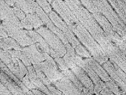
tial pattern between the periosteal and endosteal layers of bone. The second, plexiform bone,
is formed very rapidly. Since it is common for plexiform bone to develop in response to the
need for increased biomechanical support, it is often seen in larger, faster growing mammals
than humans, including cows, sheep, or deer (
Currey, 2002
). It is very rarely, if ever, seen in
humans, and if present, it will be in extremely young individuals (
Mulhern and Ubelaker,
2001
). It is characterized by rectangular, brick-like shapes (
Figure 13.2
) and stems from
mineral buds that grow first perpendicular and then parallel to the outer edge of bone surface
(
Jowsey, 1966; Martin and Burr, 1989
).
The third type, primary osteonal bone, is comprised of circular or concentric layers of
lamellae surrounding a vascular canal. These layers, the central vascular canal, and osteo-
cytes make up a primary osteon (
Jaffe, 1929
; Currey, 1982;
Martin and Burr, 1989
). In primary
bone, there is a shorter distance between osteocytes and the blood supply than in secondary
bone allowing for easier access to nutrients for growth and development (
Dempster and
Enlow, 1959
).
FIGURE 13.2
Plexiform bone.