Biology Reference
In-Depth Information
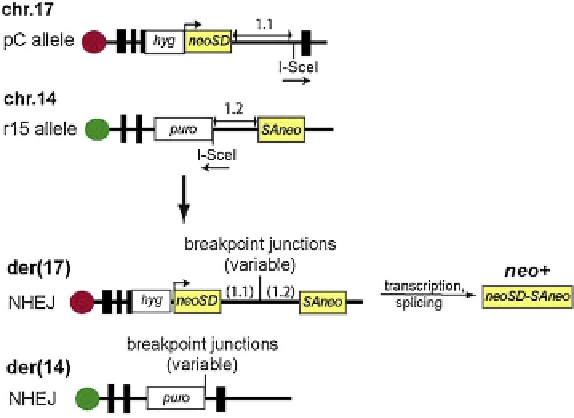
FIGURE 8.10
Reporter system for studying cellular induc-
tion of NHEJ-mediated translocations. Cells have been estab-
lished in which the two exons of the neomycin
phosphotransferase (neo) gene are located on separate chromo-
somes, 17 and 14. Each exon is fused to an intronic sequence
with an I-SceI restriction site distal to the neo exon. Cleavage of
the cellular DNA by I-SceI produces double-strand breaks with
non-palindromic overhanging termini in chromosomes 17 and
14. Translocations generated by NHEJ-mediated joining of the I-
SceI termini on different chromosomes result in derivative
chromosomes, der(17) and der(14). The former carries the two
neo exons separated by the fused introns, thereby enabling
transcription of the neo gene and rendering the cells resistant to
the drug G418. The DNA from these cells can be recovered and
sequenced between the two neo exons to analyze the newly
formed junctions. Adapted from Weinstock et al., 2006.
209
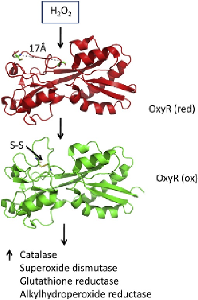
FIGURE 11.2
The response of OxyR to hydrogen peroxide is shown with ribbon representations of the reduced (top) and oxidized (bottom)
forms of the protein. In response to hydrogen peroxide, a disulfide bond forms in OxyR, changing the structure of the protein in this region, and
conferring specificity for new promoter sites in E. coli. The oxidized form of OxyR activates transcription of a number of proteins that confer
protection to the bacteria from hydrogen peroxide.