Biology Reference
In-Depth Information
Chromosomal changes, including translocations are
common features of malignant cells and it is highly
likely that NHEJ creates many of these aberrations.
207
Some of these changes can result in fusion proteins
that contribute to the carcinogenic process. For example,
the hallmark of B-cell lymphomas is the juxtaposition of
immunoglobulin loci with proto-oncogenes, resulting in
oncogene activation.
208
While it is not possible to
directly identify the sequence of events responsible for
a chromosomal translocation observed in a tumor, the
mechanism can be inferred if the cause of the DSB is
known and by examining the novel junction and deter-
mining if it is likely to match the expected sequence. In
this regard, our understanding of the formation of
translocations was greatly advanced by the elegant
in vitro
approach devised by Jasin and coworkers.
209,210
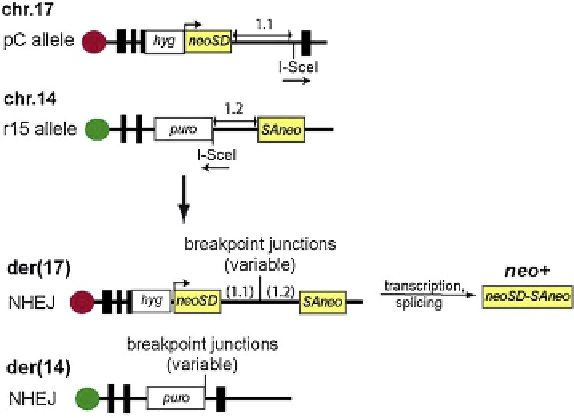
This entailed the introduction of a bipartite reporter
gene (neomycin phosphotransferase) with each exon
on a different chromosome and separated from a restric-
tion endonuclease (I-SceI) cleavage site by an intronic
sequence (
Figure 8.10
). I-SceI recognizes a non-palin-
dromic 18-bp sequence, which, when cleaved, produces
a DSB with a non-palindromic 4-base 3
0
-overhang.
Thus, following expression of I-SceI in the cells and
site-specific cleavage of the DNA, cells containing trans-
locations that arise by NHEJ-mediated joining of the
two broken chromosomes can be selected by resistance
to the drug G418 due to the splicing of the intron
between the two exons of the neomycin phosphotrans-
ferase gene and expression of the neo protein. Because
the splicing occurs in the 2.3 kb intron, a variety of
breakpoint junctions can be recovered with few
constraints other than that any deletion be less than
2.3 kb. Analysis of the breakpoint junctions showed
that 80% contained simple deletions (the majority of
which were
2% were complex. Microhomologies were found at the
vast majority of junctions. When these data were
compared to reciprocal translocations found in patient
tumor samples (primarily leukemias and lymphomas),
no significant differences were found in the percentage
of deletions or in the mean deletion length. Microho-
mologies were also common in the patient samples.
Thus based on these and other data, it was concluded
that NHEJ rather than HR is responsible for chromo-
somal translocations found in tumors.
209,210
Protective Function of NHEJ
Of course the primary function of NHEJ is to protect
cells from the consequences of DSBs. In the
in vitro
assays described above translocations between the chro-
mosomes were estimated to occur at a 1,000-fold lower
frequency than intrachromosomal DSB rejoining. The
impact of NHEJ on the tumorigenic process can be
investigated by determining the consequences of partial
or complete loss of expression of NHEJ proteins and by
examining the repair capacity of tumor and/or normal
tissues from cancer patients. Inactivating (null) or
partially inactivating (hypomorphic) mutations in
NHEJ genes have been shown to be responsible for
several rare human autosomal recessive disorders.
Elevated risk of malignancy, particularly lymphoid
cancers, is a common feature of these disorders. This
highlights the important role of incorrect DNA rejoining
during V(D)J recombination. The best-known disorder is
Ataxia telangiectasia (AT) resulting from mutations in
the
ATM
gene.
211
Clinically, AT is characterized by
progressive cerebellar ataxia (stagger when walking),
neurodegeneration, immune deficiency, radiosensitivity,
and a predisposition to cancer.
212
At the molecular level,
AT cells display cell-cycle checkpoint defects and
<
100 bp), 12% contained insertions and
FIGURE 8.10
Reporter system for studying cellular induc-
tion of NHEJ-mediated translocations. Cells have been estab-
lished in which the two exons of the neomycin
phosphotransferase (
neo
) gene are located on separate chromo-
somes, 17 and 14. Each exon is fused to an intronic sequence
with an I-SceI restriction site distal to the
neo
exon. Cleavage of
the cellular DNA by I-SceI produces double-strand breaks with
non-palindromic overhanging termini in chromosomes 17 and
14. Translocations generated by NHEJ-mediated joining of the
I-SceI termini on different chromosomes result in derivative
chromosomes, der(17) and der(14). The former carries the two
neo
exons separated by the fused introns, thereby enabling
transcription of the
neo
gene and rendering the cells resistant to
the drug G418. The DNA from these cells can be recovered and
sequenced between the two
neo
exons to analyze the newly
formed junctions. (
Please refer to color plate section
)
. Adapted from
Weinstock
et al.
, 2006.
209