Biomedical Engineering Reference
In-Depth Information
Nanosilica
20 nm
20 µm
(a)
(b)
TiO
2
pigment
Sanding
particles
from paint
matrix
TiO
2
pigment
500 nm
500 nm
(c)
(d)
FIGURE 17.1
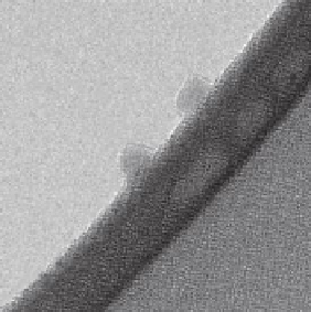
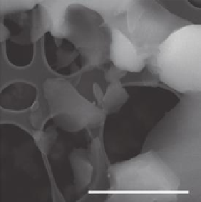
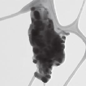
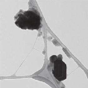
Electron microscopy images of nanosilica and different paint dusts.
(a) Transmission electron microscopy (TEM) images of Bindzil
®
CC30 nanoparticles adhered
to the edge of a hole in the carbon film of the TEM grid. The sample was prepared directly
from dispersion in in vivo test medium. (b) Scanning electron microscopy image of sanding
dust of UV-hard coat lacquer added NANOCRYL
®
XP 21/0768. The sanding dust has a wide
size distribution ranging into the nanorange, but free nanosilica particles were not observed in
the sanding dust. (c) TEM image of a paint dust particle from sanding a model reference indoor
acryl paint. TiO
2
pigments are visible inside the paint dust particle. Some occur at the surface
of the dust particle. (d) TEM image of free sub-µm-size TiO
2
pigment particles and nano- to
fine-carbonaceous particles generated during sanding the reference indoor acryl paint.
17.2.2 e
xamPles
of
n
anosiliCa
u
sed
to
a
Chieve
B
ioCidal
P
roPerties
Silica and nanosilica are not antimicrobial by themselves. However, nanocomposite
materials have been developed where nanospheres or nanoporous silica are doped
with antimicrobial or antifungal agents or even smaller nanoparticles thereof. The
potential applicability of such colloidal silica as a carrier and controlled release of
biocides such as isothiazolinone (Edge et al. 2001) as well as quaternary ammo-
nium (benzalkonium) chloride and combined silver-quaternary ammonium chloride
(Chmielewska et al. 2006; Lukasiewicz et al. 2003) have been demonstrated at least
a decade back. In both cases, the used silica materials were not described in any


Search WWH ::

Custom Search