Biomedical Engineering Reference
In-Depth Information
TABLE 9.1
Genotoxicity of Silver Nanoparticles Tested within the NanoGEM Project
Cytotoxic/Toxic to
Bacteria
Particles
Test
Result
Ag200.PVP
Micronucleus assay
in vitro
>55 µg/mL
Genotoxic > 27.5 µg/mL
Ames test
>110 µg per plate
Not mutagenic
Comet assay
Not detected up to 50 µg/cm
2
Not genotoxic
Ag50.PVP
Micronucleus assay
in vitro
>11 µg/ml
Genotoxic >11 µg/mL
Ames test
>160 µg per plate
Not mutagenic
Comet assay
Not detected up to 50 µg/cm
2
Not genotoxic
9.3 EFFECTS OF METAL NANOMATERIALS
ON ALVEOLAR MACROPHAGES
The model of primary alveolar macrophages (AM) was introduced in Chapter 8. In
the nanoGEM project, 50 nm Ag nanoparticles coated with either a polyether (Ag50.
EO, dispersed in Disperbyk 190), citrate (Ag50.citrate), or polyvinylpyrrolidone
(Ag50.PVP, dispersed in Luvitek K90) were assessed for toxic effects using AM. The
latter variant was also used as a 200 nm silver particle (Ag200.PVP). Under serum-
free cell culture conditions, Ag nanoparticles tended to incompletely agglomerate
such that dark silver grains were visible by light microscopy outside and inside AM,
a phenomenon also found to occur
in vivo
secondary to intratracheal instillation of
silver nanoparticle suspension into the lungs (Figure 9.1).
A concentration range of up to 45 µg/mL was tested. However, no “mean mass
per cell” values could be calculated because a considerable fraction of nanoparticles
remained dispersed in the supernatant. Based on the release of lactate dehydrogenase
20
µ
m
(a)
(b)
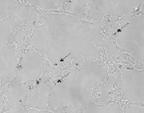
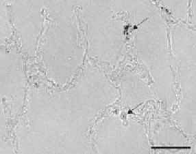
FIGURE 9.1
Bright field microscopy of unstained cryo-sections of snap-frozen rat lungs
intratracheally instilled with silver nanoparticles. The lungs were removed 30 min (a) and
3 days (b) post instillation of 0.6 mg Ag50 per lung. There is a progressive concentration of
agglomerated silver grains (arrows) in macrophage-like structures after 3 days.

Search WWH ::

Custom Search