Environmental Engineering Reference
In-Depth Information
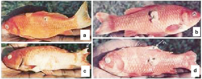
Fig. 12.6
Symptoms of
Ichthyophonus hoferi
on
Carassius carassius
(After Prabhuji and Sinha
2009
). (
a
)
Sandpaper effect and ulcerative condition. (
b
) A large
irregular hyperplastic tissue mass. (
c
) A gall on the caudal
fi n. (
d
) A gall on the dorsal fi n (
A
) and a large hyperplastic
tissue mass
has presented some quantitative data regarding
the dosage required to initiate infection. He found
that a single exposure of 50 fi sh to 2 × 10 spores
resulted in 'no infection', but several successive
exposures to the same dose on successive days
did result in infection. Using this information,
Sindermann produced an experimental epizootic
in 2000 immature Atlantic herring which resulted
in infection of 23 % of this group - 8 % acute
infections and 15 % chronic infections.
Mortalities due to acute infections occurred
within 2-4 weeks by massive invasion of the
heart and degeneration and necrosis of body mus-
culature and a minimum cellular response from
the host. The chronic phase was characterized by
a marked host cellular response leading to encap-
sulation of the parasite by fi brous connective tis-
sue. Such a condition has often been found to
exhibit pigment deposition around the spores in
the muscles.
Besides the uncoordinated swimming move-
ment of the fi sh, i.e. the swimming or reeling
movement, the affected fi sh may also exhibit
either or both the basic symptoms:
(a) Epithelioma or gall or tumour formation on
the body surface
(b) Nodulation or necrosis of vital organs like
brain, heart, liver, kidney, etc.
Signifi cant epithelioma or gall or tumour
formation has been reported (Fig.
12.6
), for the
fi rst time, by Srivastava et al. (
1984
) and Sinha
(
1985
) followed by Prabhuji and Sinha (
2009
).
These abnormal hyperplastic structures appear as
small or large tissue lumps developing on the body
surface (dorsal, lateral, peduncle region or at the
base of fi ns). On dissection the various vital organs
of the infected fi sh, viz., heart, liver, brain, kidney,
spleen, intestine and stomach, have been observed
to exhibit nodulation and necrosis (Pettit
1911
,
1913
; Rucker and Gustafson
1953
; Sindermann
and Scattergood
1954
; Dorier and Degrange
1961
;
Erickson
1965
; Srivastava et al.
1984
; Sinha
1985
;
Prabhuji et al.
1988
; Prabhuji and Sinha
2009
).
As far as control of fi sh diseases caused by
oömycetes is concerned, many chemicals have
been suggested; however, natural therapeutic
agents are rare (Prabhuji et al.
1983
,
1986
). Most
of the pure chemicals exhibit host toxicity and,
therefore, may not be used en masse. Because of
oömycetes' distinct physiology, most fungicides
are ineffective against them. With the aid of genetic
and genomic tools, oömycetes' genes encoding
secreted proteins that control the outcome of infec-
tion are being identifi ed. Ongoing genomic efforts
promise to identify further genes and create the
possibility of new control measures (Tyler
2001
).
Search WWH ::

Custom Search